Category Archives: Oral Pathology

Apert Syndrome

Apert syndrome clinical presentation

Etiology:

Genetic disorder – Premature fusion of certain skull bones (craniosynostosis) – skull does not grow normally – affects shape of head and face

Clinical:

1. Skeletal abnormalities:

  • Syndactyly
  • Acrocephaly
  • Brachicephaly

2. Oral complications:

  • High palatal vault
  • Posterior palatal cleft and bifid uvule
  • Multiple impactions and malocclusions

3. Ovoid skull + horizontal supra-orbital groove

4. Early closure of coronal & sagittal sutures

  • Mid third face underdeveloped
  • Patent alveolar & frontal sutures

Treatment:

Surgery

McCune Albright Syndrome

McCune Albright Syndrome

Etiology:

  • Mutations of Gs-α gene (guanine nucleotide binder)
  • Controls cAMP function in endocrine glands

Epidemiology:

  • Monostotic – affects 1 bone
  • Polyostotic – many bones
  • Polyostotic syndromic

Clinical:

Triad:

  1. Polyostotic fibrous dysplasia
  2. Café au lait spots on skin and mucosa
  3. Endocrine disturbances – precocious puberty

Diagnosis:

  • X-ray: Fibrous dysplasia
  • Lab: Hormone levels increase in blood test

Management:

  • Drugs to inhibit bone resorption
  • Surgery
  • Hormone therapy
  • Exercise – strengthen muscles surrounding fibrous dysplasia lesions
  • Analgesics

Achondroplasia

Achondroplasia

Etiology:

  • Dysfunction in laying down of collagen – endochondral plates in limbs not well ossified
  • AD – Replacement of ARG for GLY in gene coding FGFR3
  • Therefore constant activation of FGF
  • Therefore interference with endochondral ossification at epiphyseal growth plates of long bones

Thanatophoric dwarfism – Missense/ point mutation in FGFR3. Fatal

Clinical:

  • Long bones abnormally short
  • Skull – normal growth, trunk normal growth
  • Lordosis
  • Long trunk
  • Protrusive mandible – malocclusion

Histology:

  • Disorganized & narrow zones of proliferation & hypertrophy
  • Enlarged chondrolytes

Hereditary Craniofacial Malformations

  1. Achondroplasia
  2. Albright syndrome – McCune Albright Syndrome
  3. Apert Syndrome
  4. Behcet’s Syndrome
  5. Burning Mouth Syndrome/ Glossodynia
  6. Cleidocranial Dysplasia/ Dyostosis
  7. Cherubism
  8. Clefts of Lips & Palate
  9. Crouzon Syndrome/ Craniofacial dsyostosis
  10. Down’s Syndrome
  11. Ehlers-Danlos Syndrome
  12. Fanconi Syndrome
  13. Gardner Syndrome
  14. Gorlin Goltz Syndrome
  15. Marfan Syndrome
  16. Osteogenesis Imperfecta
  17. Osteopetrosis/ Alber’s Schonberg Disease
  18. Peutz Jegher Syndrome
  19. Pierre Robin Syndrome
  20. Plummer Vinson Syndrome
  21. Ramsay Hunt Syndrome
  22. Sjogren’s Syndrome
  23. Steven Johnson Syndrome
  24. Sturge Weber Syndrome
  25. Treacher Collins Syndrome/ Mandibulofacial Dysostosis
  26. Van der Woude Syndrome
  27. Von Recklinghausen’s Neurofibromatosis

Odontodysmorphogenesis

Atlas of dental  anomalies

1) Tooth size

1. Microdontia

Teeth measurably smaller than normal

Clinical types:

  • Absolute – smaller than normal
  • Relative – gigantism
  • Generalized – entire dentition
  • Focal – usually 7’s & 8’s

Etiology:

  • Genetic (AD) – Downs syndrome
  • Irradiation therapy of 10 Gy or more
  • Drawfism
  • Congenital heart disease
Microdontia

2. Macrodontia

Enlarged teeth

Clinical types:

  • Absolute – pituitary drawfism
  • Relative – hypognathia of maxilla/ mandible
  • Generalized – entire dentition
  • Focal – some teeth eg. hemifacial hypertrophy
Macrodontia

2) Tooth form

1. Double teeth

a) Germination – single enamel organ – 2 teeth develop

  • 2 crowns share same root canal
  • Aet: Trauma

b) Fusion – 2 tooth germs fuse by dentine ± pulp – forms single large tooth structure

  • Aet: Trauma

c) Concrescence – adjacent formed teeth join by cementum

  • Aet: Trauma, overcrowding

d) Twinning – Mirror image teeth. Complete germination

  • One tooth germ – develops into 2 teeth
Tooth anomaly - concrescence, fusion, germination

2. Dilaceration

Bending of roots at an angle at long axis of tooth

  • Aet: Trauma during root development
tooth dilaceration

3. Dens invaginatus

Abnormal exaggeration of lingual pit

  • Type I:
    • Enamel lined cavity confined to tooth crown, not extending beyond CEJ
    • Mx: Pit sealing
  • Type II:
    • Enamel lined cavity into tooth (± communication with pulp)
    • Mx: XLA
  • Type III:
    • Invagination extends beyond CEJ
    • Perforating apically or laterally at a foramen (no pulpal communication)
    • Mx: XLA
Dens invaginatus type I, II and III

4. Dens evaginatus

Anomalous tubercle/ cusp projecting from occlusal surface – usually premolar

  • Extra cusp worn down, predispose to tooth decay
Dens evaginatus

5. Taurodontism

Bull teeth

  • Elongated crowns with increased apico-occlusal height of pulp chambers
  • Failure of Hertwig’s root sheath to invaginate at proper horizontal level
Taurodontism

6. Talon cusp/ talon cingulum

Exaggeration of cusp shaped cingulum esp. Max. anteriors

  • Mx: Fissure sealing & pulpotomy
Talons cusp

7. Transposition

One tooth in place of another

Transposition

8. Supernumerary

  • Roots: Mand. canine, premolar, molar (8)
  • Cusps:
    • Cusp of carabelli
    • Enamel pearl (root furcation)
    • Dens evaginatus

3) Tooth number

1. Anodontia

Absence of teeth

  • Complete
  • Partial:
    • Hypodontia (<6 teeth)
    • Oligodontia (>6 teeth)
  • Aet:
    • 1. Failure to form dental lamina
    • 2. Failure to develop tooth buds
    • 3. Premature breakdown of dental lamina
    • 4. Autosomal dominant inheritance
Anodontia, oligodontia, hypodontia

Hereditary Ectodermal dysplasia

  • X – Linked recessive
  • Nail dystrophy
  • Hypotrichosis
  • Hyperkeratosis
  • Oligodontia

2. Pseudodontia

Clinical absence of teeth

Aet:

  • Impaction
  • Delayed eruption

3. False anodontia

Teeth exfoliated or extracted

4. Hyperdontia

Excess teeth

  • Supplemental – Normal dental series
  • Supernumerary – Abnormal morphology
  • Mesiodens – In maxillary midline
  • Paramolar/ distodens – 4th molar
  • Natal – Present at birth
  • Neonatal – Immediately after birth (30 days)
mesiodens
natal tooth

Syndromes associated with hyperdontia

1. Cleidocranial dysplasia

  • Multiple supernumeraries
  • Aplasia of one or both clavicle
  • Delayed closure of fontanelles & sutures
  • Wormian bones in suture lines
  • Short sagittal diameter of cranial base
  • Large transverse diameter of cranium

2. Gardner’s syndrome

  • Multiple unerupted supernumeraries & dentigerous cysts
  • Multiple osteomas of facial bones
  • Multiple adenomatous polyposis of colon
  • Desmoid tumors
  • Cutaneous epidermoid cyst
  • Fibrous hyperplasia of skin & mesentery

4) Tooth colour

1. Extrinsic staining

Not incorporated into tooth substance

  • Chromogenic bacteria (green, black, yellow orange)
  • Restorative materials:
    • Silver amalgam (gray black)
    • Resins (yellow brown)

2. Intrinsic staining

Incorporated into tooth structure

Changes in structure/ thickness of dental tissues:

  • Enamel hypoplasia
  • Fluorosis
  • Caries
  • Dentine dysplasia II
  • Amelogenesis imperfecta
  • Dentinogenesis imperfecta
  • Enamel opacity
  • Age changes
enamel fluorosis
Amelogenesis imperfecta

Diffusion of pigments during/ after dental development:

  • Tetracycline – cross placental barrier – yellow/ grey, brown
  • Bilirubin – green/ brown
  • Liver disease/ neonatal hepatitis – Yellow brown
  • Porphyrins
  • Pulp necrosis products
  • RH incompatibility (erythroblastosis fetalis)
  • Endodontic materials

5) Tooth eruption

1. Accelerated eruption

Symmetrical:

  • Adrogenital syndrome
  • Cerebral gigantism

Unilateral:

2. Delayed eruption

Symmetrical:

Unilateral:

  • Regional odontodysplasia
  • Ankylosing of primary predecessor
  • Dilaceration
  • Severe enamel hypoplasia:
    • Nutritional deficiancy
    • Traumatic displacement of tooth germs
  • Obstruction by impaction:
    • Rare in 1ry teeth, except 1st molar – Ankylosis/ submergence
    • Multiple impactions – Cleidocranial dysplasia
    • Single impaction:
      • Supernumeraries
      • Odontomes
      • Cysts
      • Crowding

3. Reimpaction of teeth

Previously erupted tooth becomes submerged in tissues

Aet:

  • Deficient development of alveolar process
  • Root ankylosed + Lack of growth of alveolar process

4. Premature loss

  1. Juvenile & prepubertal periodontitis
  2. Hypophosphatasia
  3. Leukemia
  4. Chronic neutropenia
  5. Acrodynia (pink disease) – Hg poisoning
  6. Dental caries
  7. Hereditary palmer – planter hyperkeratosis
  8. Langerhans cell histiocytosis – proliferation of abnormal histiocytes
    • Tooth mobility
    • Floating teeth in multiple quadrants
    • DI
    • Short stature
    • Neurosensory deafness

5. Prolonged retention

Deciduous teeth not shed

  • Mand 1’s – Lingual eruption esp. Down’s syndrome
  • Max 6’s – Path of eruption further anterior than normal

6. Occlusal defects

  1. Deep/ increased overbite:
    • Over eruption of ant. teeth
    • Infra eruption of post. teeth
  2. Open bite:
    • Usually ant. region
    • Post. mandibular growth disorder
    • Macroglossia

6) Degenerative changes (acquired)

1. Attrition

  • Physiologic wearing of teeth due to mastication
  • Abnormal occlusion habits: bruxism, tobacco/ betel chewing, abnormal tooth structure (AI, DI)
  • Histology: Reactionary dentine, dead tracts, translucent zones
attrition

2. Abrasion

  • Pathologic wear of teeth – abnormal habit, abnormal use of abrasives orally
  • Eg. Pipe smoking, tobacco chewing, aggressive tooth brushing, abrasive dentifrices, pins & nails (carpenters)
abrasion
Abrasion, abfraction, erosion

3. Erosion

  • Loss of tooth substance due to non-bacterial chemical process:
    • Occupational – car battery acid
    • Dietary – acidic drinks
    • Chronic vomiting – anorexia, bulimia
erosion

4. Resorption

  • Physiological – shed deciduous teeth
  • Pathological:
    • Periapical inflammation
    • Iatrogenic – excess orthodontic force
    • Neoplasms
    • Impacted teeth
    • Transplanted/replanted teeth
    • Idiopathic
  • External resorption: 2ry to pathology (or idiopathic)
  • Internal resorption: 2ry to pulpitis (or idiopathic)
internal resorption
Internal resorption

7) Tooth structure

A) Enamel

1. Enamel hypoplasia

  • Quantitatively defective – ameloblast fail to produce normal volume of matrix
  • Enamel normal hardness
  • Clinical:
    • Pit/ grooves on enamel surface
    • General decrease in enamel thickness
  • Ground section: Decreased prisms than normal + abnormal direction

2. Enamel hypocalcification

  • Qualitatively defective – hypomineralized
  • Normal amounts of enamel
  • Clinical:
    • White & opaque – after eruption – orange brown
    • Quickly chips off & wears off
  • Ground section: Surface layers = normal

Classification

1. Local causes
  • Trauma
  • Irradiation
  • Infection – single tooth
    • eg. Turner’s tooth – hypocalcified permanent tooth due to abscess overlying deciduous tooth (yellow/ brown)
  • Idiopathic (enamel opacities) – hypocalcification of 1ry & 2ry teeth esp. 11 and 21
2. Generalized causes
a) Environmental (chronological hypoplasia)

Birth to 6 years ie. period of crown formation of permanent teeth – ameloblast dysfunction

  • Prenatal:
    • Maternal disease
    • Rubella
    • Syphilis (T. pallidum):
      • Hutchinson’s incisors – tapered crown + notched incisal edge
      • Mulberry molars – Lobulated occlusal surfaces

NB: Disorders in calcium metabolism cause dysfunction of ameloblasts

  • Postnatal:
    • Measles
    • Chicken pox
    • Scarlet fever
    • Whooping cough
    • Pneumonia
    • Congenital heart disease
    • GIT disease
    • Hyperparathyroidism
    • Fluorosis – > 1ppm, incorporates into maturation stage (not in utero due to placental barrier)
    • Vitamin A, C, D deficiency
    • Rickets
b) Genetic

Only teeth affected: Amelogenesis imperfecta

  • Mutation on distal short arm of x chromosome – codes for amelogenin protein
  • Classification:
    • 1. Hypoplastic:
      • Areas of tooth enamel fail to form normal contour
      • Enamel is normal hardness but thin
      • Sharp needle like cusps/ pitting/ vertical grooving & wrinkling
      • Yellow + gross attrition with time
    • 2. Hypomaturation:
      • Enamel of normal thickness but wears away easily
    • 3. Hypocalcified:
      • Soft enamel lost easily after eruption
      • Yellow – brown plus gross attrition
      • Chalky consistence – Chips off

Teeth + generalized disease:

B) Dentine

1. Dentinogenesis imperfecta (hereditary opalescent dentine)

  • Autosomal dominant (AD)
  • Mutations in structural genes for collagen type 1
  • Therefore occur with osteogenesis imperfecta
  • On eruption – normal contour but opalescent amber like appearance – becomes translucent + grey/brown + bluish reflection from enamel
  • Rapidly lost attrition
  • Radiological:
    • Short, blunt roots
    • Partial/ total obliteration of pulp chamber & root canal by dentine
  • Histology:
    • Normal mantle dentine
    • Decreased tubules – wide & irregular
    • Straight DEJ (not scalloped)
    • Vascular inclusions in dentine – remnants of odontoblasts + pulp tissue
  • Biochemical:
    • Increased water content
    • Decreased mineral content
    • Decreased microhardness
  • Classification:
IIIIIIIV
ADADADAR
Opalescent dentine in 1ry & 2ryDiscoloured teethOpalescent teethBrown opalescent teeth
White scleraBlue scleraBlue/ white scleraWhite sclera
Bone fragilityBone fragility
Severe long bone deformity
Rapid dentine abrasion
Severe bone fragility
Skeletal deformity
Growth retardation
Dentinogenesis imperfecta classification

2. Shell teeth

  • Rare
  • Homozygote form of DI – No pulp chamber obliteration
  • Thin dentine, forms shell around pulp
  • Short roots

3. Dentinal dysplasia (rootless teeth)

  • Rare AD
  • Normal crown + dysplastic dentine in roots having many calcified, spherical bodies
  • Obliterated root canals & pulp chamber
  • Roots are stunted

2 types caused by fragmentation of Hertwig’s root sheath:

  1. Radicular DD:
    • Abnormal root formation
    • Periapical resorption
    • Therefore teeth exfoliate prematurely
    • X-ray: Short roots & obliterated pulp
  2. Coronal DD:
    • Discolouration of 1ry teeth
    • Obliteration of pulp chamber
    • Distorted crowns of permanent teeth
    • X-ray: Enlarged pulp chambers

NB: Metabolic disturbances affecting dentinogenesis:

  • Rickets
  • Vitamin D resistant rickets
  • Hypophosphatasia
  • Dentine dysplasia I & II

4. Regional odontodysplasia (enamel & dentine defect)

  • Involves ectodermal (enamel) & mesodermal (dentine + cementum)
  • Results from disorderly proliferation of dental epithelium at an early stage – of formation of hard tissues – in each affected
  • Etiology: Local vascular deficiency during tooth development
  • Clinical:
    • Delayed eruption
    • Irregular shape – hypoplastic and irregularly mineralized enamel
    • Dentine – thin + large interglobular dentine
    • Affects a region/ quadrant of maxilla/ mandible
    • Usually anterior maxilla and unilateral
  • X-ray:
    • Ghost teeth
    • Decreased radiopacity
    • Loss of distinction between enamel and dentine
  • Histology:
    • Abnormal odontogenic epithelial cells
    • Abnormal globular dentine
regional odontodysplasia

C) Cementum

1. Hypercementosis

Increased cementum deposition

Etiology:

  1. Root ankylosis & concrescence
  2. Periapical inflammation – Localized knob-like enlargement
  3. Mechanical stimulation – Below threshold cementum deposition, above threshold bone resorption
  4. Functionless teeth – Resorption + increased opposition
  5. Unerupted teeth – If no REE – cementum over extends surface of enamel
  6. Paget’s disease – Irregular masses and ankylosis
  7. Idiopathic

REE – Reduced enamel epithelium – lies over developing tooth

hypercementosis

2. Hypoplasia & dysplasia

  • Cleidocranial dysostosis (mentioned above)
  • Hypophosphatasia – aplasia of cementum (AR disease)

Periodontal Disease

Gingivitis

Def: Inflammation of gingiva without clinical attachment loss

Classification

1. Dental plaque induced gingival disease

  • Gingivitis associated with dental plaque only
  • Gingival disease modified by systemic factors
  • Gingival disease modified by medications
  • Gingival disease modified by malnutrition

2. Non plaque induced gingival lesion

  • Those of bacterial, viral, fungal origin
  • Genetic origin
  • Systemic origin
  • Trauma
  • Foreign body reactions

Plaque

Def: Aggregate of microorganisms – in which cells produce extracellular matrix – and are adherent to each other – and onto the tooth surface

Steps on plaque formation:

  1. Association – Dental pellicle forms
  2. Adhesion – Bacteria bind onto pellicle within hours
  3. Proliferation – of bacteria
  4. Microcolony formation
  5. Biofilm formation – Formation of symbiotic relationship
  6. Growth & maturation – Biofilm develops primitive circulatory system

Risk factors:

1. Intrinsic

  • Genetics
  • Gender
  • Race
  • Age
  • Hormones
  • Autoimmune dx
  • Diabetes
  • Hematological cancers

2. External/acquired

  • Alcohol
  • Drugs
  • Smoking
  • Stress
  • Medication use

3. Local

  • Poor oral hygiene
  • Poor restoration – overhang/defective
  • Increased calculus
  • Areas subjected to food impaction
  • Carious lesions and margins

Factors contributing to severity:

  • Pathogenic bacteria
  • Environmental factors
  • Genetic factors

Healthy gingiva:

  • Stippling gingiva, knife edge (interdental papilla), symmetrical distribution of pigment

Periodontitis

Def: Inflammation of supporting tissues of the teeth with clinical attachment loss

Clinical classification

1. Prepubertal (accelerated) periodontitis – Rare, genetic/medical condition

2. Juvenile Periodontitis – Uncommon, puberty & adolescence

  • More in females
  • Clinical presentation – Rapid destruction of alveolar bone. Starts with central incisors & first molars
  • Etiology: Bacteria
    • Actinobacillus actinomycetemcomitans
    • Capnocytophagea
    • Eikenella corrodens

3. Rapidly progressive/ aggressive – Uncommon, late adolescence

  • From puberty to 30 years of age
  • Affects entire dentition (1ry and 2ry)
  • Etiology: Leukocyte dysfunction

4. Chronic adult periodontitis – Common, adults over 30

  • Chronic bacterial destruction + plaque + calculus deposits

Chronic adult periodontitis

– Evidence of CAP is of bacterial origin:

  1. Epidemiological studies – +ve association between dental plaque & PDL disease
  2. Clinical experiment – No oral hygiene – plaque and gingivitis present
  3. Topical antimicrobials eliminate disease
  4. Isolate pathological bacteria from bacterial pocket
  5. Bacteria from plague cause disease in gnotobiots

– Systemic factors that cause disease progression:

  1. DM
  2. Pregnancy – Estrogen, progesterone
  3. Nutrition – Protein & Vit C deficiency
  4. Smoking – impaired PMN function
  5. AIDS
  6. Blood disease – Leukemia:
    • General gingival enlargement
    • Gingival bleeding
    • Gingival ulceration
    • Necrotizing ulceration
    • Mucosal pallor
    • Petechial hemorrhage
  7. Drugs:
    • Antileptics* – Phenytoin
    • Immunosuppressants* – Cyclosporin, Azathioprine, Corticosteroid
    • Calcium channel blockers* – Nifedipine, Verapamil
    • NSAIDs – Ibuprofen, Indomethacin
    • Sex hormones – Estrogen, progesterone (exacerbate gingivitis)

*Cause gingival hyperplasia

– Clinical stages in CAP

Healthy gingiva Chronic gingivitisChronic periodontitis
Aerobes : Fac. anaerobes75 : 2560 : 4020 : 80
G+ve : G-ve90 : 1065 : 3525 : 75
Motile : Non motile1 : 40↑ motility rods & spirochetesAbundant motile rods & spirochetes
Main speciesStreptococcus
Actinomyces
Veilonella
S, A, V
Capnocytophaga
Fusobacteria
Prevotella
Porphyromonas
A, F, P, P
Camphylobacter
Eikenella

Pathogenesis of PDL disease

Progression depends on balance between host & microbial factors

Host factors:

  1. Salivary factors
  2. Epithelial barrier
  3. Crevicular fluid
  4. Immune response
  5. Transmigrating neutrophils
  6. Tissue regeneration & repair

Microbial factors:

  1. Direct injury
  2. Toxic products
  3. Enzymes
  4. Antigenic challenge

Disturbance in host – parasite relationship leads to:

  1. Activation of host immune & inflammatory response
  2. ↑ Synthesis of inflammatory mediators & cytokines (IL-1, IL-6)
  3. Breakdown of CT and bone resorption
  4. Progression of PDL disease

Clinical progression of PDL disease

1. Initial lesion: 2-4 days, base of gingival sulcus. Gingivitis

  • Acute inflammation:
    • Vasodilation
    • Crevicular fluid exudates
    • PMN infiltrates
    • Neutrophil and monocytes appear
  • Disruption of junctional epithelium

2. Early lesion: 4-7 days, exacerbation of initial gingivitis

  • Change in junctional epithelium:
    • Disruption of intercellular spaces
    • Detachment from enamel
    • Deepening of gingival sulcus
    • Subgingival plaque formation
  • Lymphocytic infiltrate
  • Loss of collagen
  • Hyperplastic junctional epithelium (JE)
  • Damage fibroblast cell membrane & organelles

3. Established lesion: 2-3 weeks, disrupt JE. Gingivitis

  • Ulceration of gingival pocket epithelium – plasma cell infiltrate
  • Destruction of gingival CT – expansion of involved tissues
  • Hyperplastic gingivitis (repair)

4. Advanced lesion: Periodontitis – 3 weeks – inflammation spreads to alveolar bone & PDL

  • Destruction of CT:
    • Collagen degradation
    • Bone resorption
    • Pocket formation
  • 50% plasma cells
  • Clinical attachment loss (CAL) > 3mm

– Mechanism of degradation of CT and collagen:

  1. Cytopathic changes in fibroblasts – ↓ synthetic activity
  2. ↑ enzyme activity of:
    • Collagenase (produced by bacteria & fibroblast)
    • Metalloproteinases (inflam. cytokines)
    • Other proteases

– Pathological features of established PDL disease:

  1. Persistent gingivitis
  2. Loss of CT attachment
  3. Destruction of alveolar bone
  4. Predominance of plasma cells
  5. Apical extension of destructive inflammation
  6. Healing with periods of quiescence

Long term effects of apical periodontitis

  • Apical periodontitis: Tender on mastication, edema and inflammation in pdl. Widened pdl due to bone resorption in x-ray.

Other periodontal conditions

1. Lateral PDL abscess

Localized area of pus in PDL pocket

  • Acute:
    • Develops rapidly
    • Redness, swelling, tenderness of overlying mucosa
    • Throbbing pain
  • Chronic – dull pain/ asymptomatic

Picture

2. Pericoronitis

  • Pain
  • Bad taste
  • Pus discharge
  • Tender gum flap

Acute pericoronitis – Common site – 7 & 8

  • Pain
  • Trismus (lock jaw)
  • Fever
  • Hyperemia
  • lymphadenitis
  • Swelling
  • Halitosis
  • Tx: Antibiotics, operculectomy, disimpaction

Picture

3. Acute necrotizing gingivitis

Disease of young people

Etiology:

  • Borrelia vincentii
  • Fusiform bascilli

Clinical presentation:

  • Interdental papillae ulcers
  • Gingival bleeding
  • Halitosis
  • Sialorrhoea (drooling)

Predisposing feature:

  • Respiratory infection
  • Poor oral hygiene
  • Smoking
  • Immunodeficiency & HIV
  • Malnutrition

Picture

4. NOMA/ Cancrum oris/ Gangrenous stomitis

  • Rapidly progressive, polymicrobial, opportunistic infection – leads to destruction of orofacial tissues
  • Malnourished children

Etiology:

  • Borrelia vincentii
  • Staph. aureus
  • Prevotella intermedia
  • Fusobacterium necrophorum

Underlying systematic disease:

  • Malnutrition, poverty
  • Dehydration
  • Poor oral hygiene
  • Burkitt’s lymphoma
  • HIV
  • Leukemia
  • Noma neonatorum – low birth weight, premature ill infants
  • Debilitating childhood disease
  • Pneumonia
  • Sepsis
  • Oral mucosal ulcers
  • Trauma
  • Acute necrotizing gingivitis – antecedent lesion

Clinical presentation:

  • Tissue necrosis
  • Painful ulcers on gingiva/ buccal mucosa
  • Rapidly spreading – cause necrosis
  • Fetid odor
  • Exfoliated teeth
  • Sequestration

Complication:

  • Sequestration of involved bone
  • Soft tissue necrosis
  • Keloid formation
  • Microstomia and micrognathia

Management:

  • Treat systemic disease
  • Rehydrate
  • Antibiotics
  • Debridement of necrotic tissue
  • Reconstructive surgery

Picture


Generalized enlargement of gingiva:

  1. Local factors – Plaque, calculus, bacteria
  2. Hormonal imbalance – Estrogen, progesterone, vit C deficiency
    • Pregnancy, puberty, pills
  3. Systemic disease – Leukemia, Wegener’s granulomatosis
  4. Genetic factors – Cowden syndrome (multiple oral papilloma)
  5. Drugs – Phenytoin, cyclosporin, nifedipine, verapamil
  6. Gingival fibromatosis
  7. Chronic hyperplastic gingivitis

Clinical presentation:

  • Increased bulk of gingiva
  • Loss of stippling
  • Gingival margins rolled & blunted
  • Red-blue if inflamed

Histology:

  • Abundant collagen
  • Scanty fibroblasts
  • Inflammatory infiltrate
  • Leukemia – atypical & immature WBC

Management:

  • Gingivectomy/ gingivoplasty
  • Prophylaxis
  • OHI (Oral hygiene instructions)

Picture

Oral Infection – Acute and Chronic

Acute Infections

1. Periapical osteitis – Localized inflammation of bone marrow spaces. Throbbing pain and tenderness to vertical percussion.

2. Phoenix abscess – Acute inflammation superimposed on a chronic lesion eg. cyst or granuloma

3. Pericoronitis – Infection of soft tissues (operculum) surrounding the crown of a partially erupted tooth

4. Folliculitis – Infection of follicle of developing 2ry teeth (spreads from 1ry predecessors)

5. Fistula – Abscess communicates with an epithelial surface

6. Ludwig’s angina – Rapidly spreading – septic cellulitis – involving submental, submandibular & sublingual spaces bilaterally

NB:

Primary fascial spaces: Adjacent to origin of infection

  • Vestibular
  • Sublingual, sub mandibular, submental
  • Canine
  • Buccal

Secondary fascial spaces:

  • Pterygomandibular space
  • Masseteric
  • Superficial and deep temporal
  • Infratemporal
  • Masicator
  • Lateral pharyngeal
  • Retropharyngeal
  • Prevertebral
AbscessCellulitis
Localized collection of pusInflammation of CT, non suppurative
Pocket with necrotic tissue, bacterial colonies and dead white cellsWarm, diffuse, erythematous, indurated tissue in infected area
ChronicAcute
Localized painSevere and generalized pain
Well circumscribedDiffuse
FluctuantIndurated
AnaerobicAerobic
Difference between abscess and cellulitis

Chronic Infections

1. Chronic dento-alveolar abscess – Abscess from dental tissues spreads into alveolar bone. Sinus through alveolar bone onto mucosa near level of apex. Tender on percussion, no EPT response, Ice relieves pain, heat aggravates. Management: Drainage via pulp chamber/trephination, irrigate with H2O2 and normal saline, antibiotics

2. Condensing osteitis – Deposition of bone along existing trabeculae due to chronic irritation

3. Osteosclerosis – Deposit compact bone within trabecular area

4. Granuloma – mass of granulation tissue, consists of:

  • Proliferating capillaries
  • Fibroblasts
  • Lymphocytes
  • Plasma cells
  • Macrophages
  • Giant cells
  • Collagen deposits – more pronounced at periphery

5. Periapical scar – Dense fibrous tissue. Scar forms after periapical inflammation resolves

6. Periostitis – Inflammation of periosteum (Vascular CT enveloping bones)

7. Hypercementosis – Excessive deposition of cementum

8. Osteomyelitis – Bone marrow infection


Potentially fatal complications of oral infections

1. Intracanal spread by septic emboli

Cavernous sinus and pterygoid plexus communication. Foramen ovale, foramen lacerum

2. Mediastinitis

3. Bacteremia – detectable levels of bacteria in blood

4. Septicemia – Increased bacteria + toxins in blood

5. Septic shock – Septicemia, G-ve bacteria, inadequate perfusion of tissues

6. Necrotizing fasciitis


In summary:

Untreated dental infections can lead to:

A) Local:

  • Periapical abscess/granuloma
  • Root resorption
  • Cellulitis eg. Ludwig’s angina
  • Osteomyelitis
  • Osteosclerosis
  • Trismus

B) Systemic (ascending):

  • Cavernous sinus thrombosis
  • Meningitis
  • Extrusion of orbit

C) Systemic (descending):

  • Mediastinitis
  • Bacteremia
  • Septicemia
  • Septic shock
  • Necrotising fasciitis

Cariology – Study of Dental Caries

Def: A bacterial disease – of calcified tissues of teeth – involving demineralization of the inorganic structure – and destruction of organic substance – of the tooth.

Etiology of caries

1. Cariogenic bacteria:

  • S. Mutans
  • S. Sobrinus
  • S. Oralis
  • S. Sanguis
  • S. Mitis
  • Lactobacilli
  • Actinomyces

2. Susceptible tooth surface

3. Substrate

4. Time

NB: Acidogenic Theory: W. D. Miller 1889

  • Oral bacteria – ferment dietary CHO – form acid – decalcify tooth substance + destroy organic matrix


Experimental evidence: Role of S. Mutans in caries

1. Rapid generation of acid from sucrose

2. Synthesize extracellular polysaccharides

  • To promote adhesion to tooth
  • Increase plaque bulk

3. Synthesize intracellular polysaccharides – sustains acid production in the absence of sucrose

4. Present in high numbers in plaque associated lesions

5. Cariogenic and respond to immunizations in animal models

NB: This evidence is based on Koch’s postulates (1877)

  • 1. Isolate organism from pt with dx
  • 2. Culture organism outside the body
  • 3. Organism can cause dx in healthy susceptible animals
  • 4. Recover organism from inoculated animal. Pt’s serum contains antibodies to the organism.

NB:

  • Gnotobites + Cariogenic diet: No caries
  • Gnotobites + Cariogenic diet + Inoculated cariogenic bacteria: Caries develop

Etiology variables for caries progression

Intrinsic factors

Advantageous:

  • Increased flouride conc. in enamel
  • Decreased enamel solubility

Disadvantageous:

  • Enamel hypoplasia and hypomineralization
  • Deep pits and fissures
  • Misaligned teeth

Extrinsic factors

1. Saliva

  • Flow rate – High flow rate, decreased caries
  • Viscosity – Low viscosity, increased caries
  • Buffer (Ca2+ and Po42-)
  • Antimicrobial agents – Immunoglobulins, lysozyme, lactoferrous, thiocyanates

2. Diet

  • Presence of phosphates – Decreases caries
  • Fat – Increases caries
  • Trace elements (molybdenum, vanadium) – Decrease caries

Caries classification

1. Site of attack

  • Pit/fissure caries
  • Smooth surface caries
  • Cemental/root caries
  • Recurrent caries

2. Rate of attack

  • Rampant/acute caries
  • Slow/chronic caries
  • Arrested caries

Factors affecting outcome of caries

1. Nature of irritant – Bacterial type and load

2. Duration and severity of irritant – Bacterial virulence

3. Amount of bacterial substrate

4. Apical blood flow

5. Local anatomy of pulp chamber

6. Pre-existing state of pulp

7. Extent of other trauma

8. Microbial factors – Pathogenicity is the ability of a microbial species to produce dx = Virulence

  • Adhesion – Bacteria to tooth surface
  • Invasiveness – Spread to host tissue after infection
  • Toxigenecity – Endotoxin (cell wall), Exotoxin (excrete)
  • Communicability – Spread from one host to another

9. Host defenses

– Local

  • Epithelial lining – Physical barrier, IgA
  • Saliva
  • Colonization by normal flora

– Systemic

  • Humoral immunity – Ig A, D, E, M, G
  • Complement system & cytokines
  • Cell immunity – Lymphocytes (B & T), PMN’s

Spread of microbes from dento-alveolar complex

  1. Direct invasion/extension
  2. Lymphatic
  3. Haematogenous

Histopathology of enamel caries

Ground sections of teeth examined by:

  • Transmitted light
  • Polarized light
  • Microradiography
  • Electron microscopy (Microdissected pieces of enamel)
  • Biochemical studies (Microdissected pieces of enamel)

Phases of enamel caries:

  • The early (submicroscopic) lesion
  • Phase of non bacterial enamel crystal destruction
  • Cavity formation
  • Bacterial invasion of enamel
  • Undermining of enamel from below after spread into dentine

Histological Zones:

Histological zones of enamel caries

NB: Due to absence of cellular sensors in enamel – there is no defense reaction

1. Surface zone: 40 um thick

  • Little change in early lesion
  • Highly mineralized: Fluoride and Magnesium
  • Increased proteins
  • Surface of enamel is resistant to acid attack due to its structure, therefore subsurface demineralization occurs
  • Reprecipitation of minerals from:
    • Plaque
    • Minerals dissolved from deeper layers of lesion

2. Body of lesion: 2-25% pore volume

  • Reprecipitation of minerals (from deeper zones) – form larger apatite crystals
  • Continuous acid attack – more destruction of minerals – replaced by unbound water and organic matter from saliva and microbes
  • Increased prominence of striae of Retzius: White spots

3. Dark zone: 2-4% pore volume

  • Some remineralization concurrent with destruction
    • Narrow zone: Rapidly advancing lesion
    • Wide zone: Slow advancing lesion
  • Reprecipitation occurs from translucent zone

4. Translucent zone: 1% pore volume = size of H2O molecule

  • Normal enamel has 0.1% pore volume, therefore more porous than normal enamel
  • 1st recognizable change at advancing edge of lesion
  • Selective dissolution of Mg2+ and CO3-2 – Occurs at junctional areas btwn prismatic and interprismatic areas

NB: White spots stained with exogenous pigments (food, tobacco, bacteria) become brown spots


Histopathology of dentine caries

Defense reaction mediated by pulpo-dentinal complex

  • Sclerosis
  • Reactionary dentine
  • Sealing of dead tracts

Caries destruction involves interacting processes

  • Demineralization (acid always in advance of bacteria)
  • Proteolysis of matrix

NB: Defense reaction of dentine may occur before caries reach dentine due to irritation of odontoblasts – tertiary dentine

NB: Circumpulpal dentine more resistant to carious attack due to increased F diffused into from pulp

Histopathology of dentine caries

1. Zone of sclerosis (Translucent zone)

  • Sclerosed dentine = Increased mineral content
  • 2 patterns of mineralization:
    • 1) Centripetal deposition of peritubular dentine – therefore occlude tubule
    • 2) Minerals appear in cytoplasmic process of odontoblast – tubule occluded by odontoblastic processes
  • Dead tracts: Opaque, seen in sclerotic zone
    • Empty tubules + air + degraded odontoblastic processes
    • Occluded at pulpal end of EBURNOID (thin layer of hyaline calcified material)

2. Zone of demineralization

  • Wave of acid from bacteria in this zone
  • No bacteria in this zone

3. Zone of bacterial invasion

  • Bacteria multiply and extend within tubule
  • Acidogenic organisms eg. lactobacilli – occupy tubules at periphery of lesion and produce acids
  • Acidogenic + proteolytic organisms – attack demineralized matrix, therefore soften walls of tubules

Increased bacteria + Compression of inter and peritubular dentine = Break down of dentinal tubules – Liquefaction area (elliptical area)

The liquefaction areas are called liquefaction foci – which enlarge, increase in number and coalesce

Liquefaction area/foci dentinal tubules

4. Zone of destruction

  • Cavitation at amelodentinal junction
  • Transverse clefts (cracks) appear in dentine – perpendicular to the direction of dentinal tubules
  • Contain bacteria and necrotic tissue
  • Bacteria invade peri and intertubular dentine

5. Reactionary/Tertiary dentine

  • Beneath lesion in pulp
  • Pre-odontoblasts differentiate – lay down tertiary dentine to protect pulp

Histopathology of cementum caries

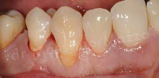
  • Recession of gingiva – cementum exposed to oral environment – cementum more homogenous and laminated – therefore when attacked – caries produce saucer shaped lesions
  • Prominent microbes:
    • Strep. mutans
    • Actinomycosis viscosus

Diagnostic Methods

A) Imaging Techniques

1. Plain Radiograph

2. Contrast Radiograph – Radiopaque substance introduced in body

Sialography:

– Iodine

Angiography

  • Fluorescent dye/fluoroscope
  • Catheter introduced into artery (external carotid)

TMJ arthroscopy

  • Patient discomfort
  • Contrast introduced in joint space

Radionuclide imaging

  • Technetium 99 – injected into blood stream – labelled with methylene diphosphonate (MDP) – affinity for bone
  • Indications:
    • Bone metastasis
    • Assess bone graft
    • Assess condylar growth
    • Assess thyroid function
    • Investigate salivary glands
  • Disadvantages:
    • High dose needed
    • Poor resolution
    • Non specificity

3. Digital Imaging – Receptor converts x-ray image into digital data and stores on computer

  • Receptor:
    • Charged couple device (CCD)
    • Photo stimulable sensor plate
  • Advantages:
    • Low radiation dose
    • No conventional processing
    • Manipulate image with software
    • Efficient record keeping
  • Disadvantages:
    • Expensive
    • Bulky sensor for intraoral space
    • Low resolution compared to x-ray film
    • Misuse of image manipulation
    • Occupy a lot of disc space

4. Conventional Tomography – Greater range of movement, image tissues in slices

5. Computed Tomography (CT Scan)

  • Radiographic tube passes in a circle around the body – detectors measure degree of attenuation (blocking) by tissues – value converted to numerical value (Hounsfield number) – digitally converted to graphic image
  • Indications:
    • Mid facial trauma
    • Bone dx

6. MRI

  • Patient in strong magnetic field – pulses of radiowaves – impart energy – hydrogen protons in body fluid – emit radiosignal – detected and processed by computer
  • Has no ionizing radiation (compared to CT or xray)
  • Types:
    • T1 – Highlights fat tissue eg. subcutaneous fate, bone marrow
    • T2 – Highlight water eg. CSF
  • Indications:
    • Soft tissue detail
    • Tumour staging
    • Intracranial dx
    • TMJ (except bone) lesions
  • C/I:
    • Pt with pacemaker
    • Ear implant
    • 1/3 of pregnancy
    • Ferromagnetic surgical clips
    • Bone lesions

7. Positron Emission Tomography (PET) – Radiation/nuclear medicine to produce 3D colour image

8. Ultrasonography

  • Indication – Superficial soft tissue eg. Salivary gland, thyroid

B) Specimens for Lab Investigations

1. Haematology

EDTA:

  • Cross matching
  • All red cell indices
  • White cell counts
  • Platelet counts
  • Serum vit B12
  • Blood films
  • RBC folate assays

Plain Tube:

  • Cross matching
  • Blood grouping
  • Serum iron
  • Serum ferritin
  • TIBC

Citrated Tube:

  • ESR
  • Prothrombin time

2. Biopsy – Histopathology

  • 10x volume of biopsy needed
  • Fixed in 10% solution of formal saline
  • Immediate freezing at -70 degrees Celsius for immunofluorescent exam

3. Microbiology – Culture and sensitivity

  • Collect before antimicrobe tx – if cannot process in 2 hours – place swab in transport media and store at 4 degree Celcius

NB: Don’t take samples for viral hepatitis and HIV, refer to VCT clinic


C) Biopsy Techniques

  1. Excisional – Entire lesion + margin of healthy tissues (benign lesions)
  2. Incisional – Portion of lesion + margin of healthy tissues
  3. Punch – Needle removes small portion (tumors with high risk of seeding)
  4. Smear/brush – Exfoliative cytology (superficial lesion on oral mucosa)
  5. Aspiration – Wide bore needle into lesion and aspirate (cysts)

D) Microscopic Techniques

1. Light Microscopy

  • Bright field/standard: Wet films – bacterial motility
  • Darkground microscopy: Dental ground sections – illuminate obliquely
  • Phase – contrast: Details of unstained microbes
  • Flourescent microscopy: Immunology

2. Electron Microscopy: Resolution of small microbes (virology)


E) Lab Isolation of Microbes

1. Bacteria

Stains:

a) Gram Stain – Lugol’s iodine, carbolfuchsin

b) Ziehl – Nielsen – Carbolfuchsin + methylene blue/ malachite green

  • Mycobacteria (thick waxy wall)
  • +ve: Red against blue background

Culture media:

a) MacConkey

  • Red fermenters – E.coli, Kleibsiella, Citrobacter, Enterobacter
  • Yellow/ non fermenters – Salmonella, Pseudomonas

b) Mitis Salivarius

  • > 2mm – Strep. Salivarius
  • < 1mm – Strep. Mitis

c) Mannitol Salt

  • Big, yellow – Staph. Aureus
  • Small, pink – Staph. Epidermidis

d) Lowenstein – Jensen

  • Rough – M. Tuberculosis
  • Smooth – Atypical Mycobacteria

e) TCBS (Thiosulphate, citrate, bile, saliva)

  • Yellow fermenters – V. Cholerae, Acromonas
  • Non Fermenters – V. Parahaemolyticus

f) Thayer Martin

  • Grey colonies – Neisseria

g) Charcoal Yeast

  • Cutglass colonies – Legionella

Universal transport media

Stuart transport media (semi – solid, non nutrient agar)

2. Fungi

Stains:

  • Periodic Acid Schiff
  • Gram stain
  • Methenamine silver

Culture media

  • Sabouraud’s agar
  • Blood agar
  • Cornmeal agar (cream colonies)

Identification test: Germ Tube Test

  • Unknown candida spp + Serum incubation for 3 hours at 37°C
  • +ve: Growth of new hyphae (germ tubes)
  • -ve: Persistance of candidal spores

Transport media

  • Nutrient agar/Broth

3. Viruses

Polymerase chain reaction

Smear biopsy

  • Examine cells for degenerative changes eg. rounding of epithelial cells, cell fusion, multinucleate cells

Serodiagnosis

  • ELISA
  • Immunoflourescence – Direct and indirect
  • Rising antibody titers
Direct and indirect flourescence

4. Parasites

Stains

  • Giemsa stain – Malaria, Leishmania
  • Periodic Acid Schiff stain – Amoebae
  • Silver stain – Pneumocystis