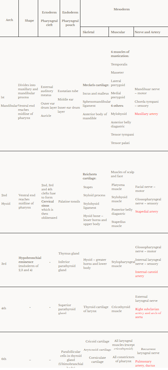
6 mesodermal thickenings on each side of primitive pharynx:
- Outer ectodermal covering (Externally – 5 grooves – Pharyngeal clefts)
- Mesodermal core
- Inner endodermal lining (Internally – 4 grooves – Pharyngeal pouches)


Basic structure of pharyngeal arch:
- Mesoderm – Cartilagenous bar (forms cartilage, ligaments and bones)
- Mesoderm – Striated muscle (special viceral muscle of head and neck)
- Aortic arch artery
- Own nerve (motor + posttrematic sensory)
- Next arch’s nerve (pretrematic sensory)
NB: 5th arch disappears
Derivatives of the arches:
Ectoderm, endoderm, mesoderm derivatives and shape of each pharyngeal arch:
Anomalies:
- Branchial cyst – along anterior border of sternocleidomastoid muscle due to failure of cervical sinus to obliterate
- Branchial sinus – Branchial cyst opens into skin by narrow canal
- Branchial fistula – Branchial cyst opens into lumen of pharynx
- Duplication of external auditory meatus – 1st branchial anomaly
Note: If left untreated, may become repeatedly infected and inflamed. Recurrent inflammation makes surgical resection more difficult. Excellent prognosis if lesion is completely resected.
