A) Imaging Techniques
1. Plain Radiograph
2. Contrast Radiograph – Radiopaque substance introduced in body
Sialography:
– Iodine
- Ductal system of parotid and submandibular glands
- Indications: Ductal obstruction, Sjogrens syndrome
- Contra-indications: Acute sialadenitis, Salivary masses
Angiography
- Fluorescent dye/fluoroscope
- Catheter introduced into artery (external carotid)
TMJ arthroscopy
- Patient discomfort
- Contrast introduced in joint space
Radionuclide imaging
- Technetium 99 – injected into blood stream – labelled with methylene diphosphonate (MDP) – affinity for bone
- Indications:
- Bone metastasis
- Assess bone graft
- Assess condylar growth
- Assess thyroid function
- Investigate salivary glands
- Disadvantages:
- High dose needed
- Poor resolution
- Non specificity
3. Digital Imaging – Receptor converts x-ray image into digital data and stores on computer
- Receptor:
- Charged couple device (CCD)
- Photo stimulable sensor plate
- Advantages:
- Low radiation dose
- No conventional processing
- Manipulate image with software
- Efficient record keeping
- Disadvantages:
- Expensive
- Bulky sensor for intraoral space
- Low resolution compared to x-ray film
- Misuse of image manipulation
- Occupy a lot of disc space
4. Conventional Tomography – Greater range of movement, image tissues in slices
5. Computed Tomography (CT Scan)
- Radiographic tube passes in a circle around the body – detectors measure degree of attenuation (blocking) by tissues – value converted to numerical value (Hounsfield number) – digitally converted to graphic image
- Indications:
- Mid facial trauma
- Bone dx
6. MRI
- Patient in strong magnetic field – pulses of radiowaves – impart energy – hydrogen protons in body fluid – emit radiosignal – detected and processed by computer
- Has no ionizing radiation (compared to CT or xray)
- Types:
- T1 – Highlights fat tissue eg. subcutaneous fate, bone marrow
- T2 – Highlight water eg. CSF
- Indications:
- Soft tissue detail
- Tumour staging
- Intracranial dx
- TMJ (except bone) lesions
- C/I:
- Pt with pacemaker
- Ear implant
- 1/3 of pregnancy
- Ferromagnetic surgical clips
- Bone lesions
7. Positron Emission Tomography (PET) – Radiation/nuclear medicine to produce 3D colour image
8. Ultrasonography
- Indication – Superficial soft tissue eg. Salivary gland, thyroid
B) Specimens for Lab Investigations
1. Haematology
EDTA:
- Cross matching
- All red cell indices
- White cell counts
- Platelet counts
- Serum vit B12
- Blood films
- RBC folate assays
Plain Tube:
- Cross matching
- Blood grouping
- Serum iron
- Serum ferritin
- TIBC
Citrated Tube:
- ESR
- Prothrombin time
2. Biopsy – Histopathology
- 10x volume of biopsy needed
- Fixed in 10% solution of formal saline
- Immediate freezing at -70 degrees Celsius for immunofluorescent exam
3. Microbiology – Culture and sensitivity
- Collect before antimicrobe tx – if cannot process in 2 hours – place swab in transport media and store at 4 degree Celcius
NB: Don’t take samples for viral hepatitis and HIV, refer to VCT clinic
C) Biopsy Techniques
- Excisional – Entire lesion + margin of healthy tissues (benign lesions)
- Incisional – Portion of lesion + margin of healthy tissues
- Punch – Needle removes small portion (tumors with high risk of seeding)
- Smear/brush – Exfoliative cytology (superficial lesion on oral mucosa)
- Aspiration – Wide bore needle into lesion and aspirate (cysts)
D) Microscopic Techniques
1. Light Microscopy
- Bright field/standard: Wet films – bacterial motility
- Darkground microscopy: Dental ground sections – illuminate obliquely
- Phase – contrast: Details of unstained microbes
- Flourescent microscopy: Immunology
2. Electron Microscopy: Resolution of small microbes (virology)
E) Lab Isolation of Microbes
1. Bacteria
Stains:
a) Gram Stain – Lugol’s iodine, carbolfuchsin
b) Ziehl – Nielsen – Carbolfuchsin + methylene blue/ malachite green
- Mycobacteria (thick waxy wall)
- +ve: Red against blue background
Culture media:
a) MacConkey
- Red fermenters – E.coli, Kleibsiella, Citrobacter, Enterobacter
- Yellow/ non fermenters – Salmonella, Pseudomonas
b) Mitis Salivarius
- > 2mm – Strep. Salivarius
- < 1mm – Strep. Mitis
c) Mannitol Salt
- Big, yellow – Staph. Aureus
- Small, pink – Staph. Epidermidis
d) Lowenstein – Jensen
- Rough – M. Tuberculosis
- Smooth – Atypical Mycobacteria
e) TCBS (Thiosulphate, citrate, bile, saliva)
- Yellow fermenters – V. Cholerae, Acromonas
- Non Fermenters – V. Parahaemolyticus
f) Thayer Martin
- Grey colonies – Neisseria
g) Charcoal Yeast
- Cutglass colonies – Legionella
Universal transport media
Stuart transport media (semi – solid, non nutrient agar)
2. Fungi
Stains:
- Periodic Acid Schiff
- Gram stain
- Methenamine silver
Culture media
- Sabouraud’s agar
- Blood agar
- Cornmeal agar (cream colonies)
Identification test: Germ Tube Test
- Unknown candida spp + Serum incubation for 3 hours at 37°C
- +ve: Growth of new hyphae (germ tubes)
- -ve: Persistance of candidal spores
Transport media
- Nutrient agar/Broth
3. Viruses
Polymerase chain reaction
Smear biopsy
- Examine cells for degenerative changes eg. rounding of epithelial cells, cell fusion, multinucleate cells
Serodiagnosis
- ELISA
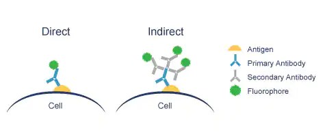
- Immunoflourescence – Direct and indirect
- Rising antibody titers
4. Parasites
Stains
- Giemsa stain – Malaria, Leishmania
- Periodic Acid Schiff stain – Amoebae
- Silver stain – Pneumocystis
