Acute Infections
1. Periapical osteitis – Localized inflammation of bone marrow spaces. Throbbing pain and tenderness to vertical percussion.
2. Phoenix abscess – Acute inflammation superimposed on a chronic lesion eg. cyst or granuloma
3. Pericoronitis – Infection of soft tissues (operculum) surrounding the crown of a partially erupted tooth
4. Folliculitis – Infection of follicle of developing 2ry teeth (spreads from 1ry predecessors)
5. Fistula – Abscess communicates with an epithelial surface
6. Ludwig’s angina – Rapidly spreading – septic cellulitis – involving submental, submandibular & sublingual spaces bilaterally
NB:
Primary fascial spaces: Adjacent to origin of infection
- Vestibular
- Sublingual, sub mandibular, submental
- Canine
- Buccal
Secondary fascial spaces:
- Pterygomandibular space
- Masseteric
- Superficial and deep temporal
- Infratemporal
- Masicator
- Lateral pharyngeal
- Retropharyngeal
- Prevertebral
| Abscess | Cellulitis |
| Localized collection of pus | Inflammation of CT, non suppurative |
| Pocket with necrotic tissue, bacterial colonies and dead white cells | Warm, diffuse, erythematous, indurated tissue in infected area |
| Chronic | Acute |
| Localized pain | Severe and generalized pain |
| Well circumscribed | Diffuse |
| Fluctuant | Indurated |
| Anaerobic | Aerobic |
Chronic Infections
1. Chronic dento-alveolar abscess – Abscess from dental tissues spreads into alveolar bone. Sinus through alveolar bone onto mucosa near level of apex. Tender on percussion, no EPT response, Ice relieves pain, heat aggravates. Management: Drainage via pulp chamber/trephination, irrigate with H2O2 and normal saline, antibiotics
2. Condensing osteitis – Deposition of bone along existing trabeculae due to chronic irritation
3. Osteosclerosis – Deposit compact bone within trabecular area
4. Granuloma – mass of granulation tissue, consists of:
- Proliferating capillaries
- Fibroblasts
- Lymphocytes
- Plasma cells
- Macrophages
- Giant cells
- Collagen deposits – more pronounced at periphery
5. Periapical scar – Dense fibrous tissue. Scar forms after periapical inflammation resolves
6. Periostitis – Inflammation of periosteum (Vascular CT enveloping bones)
7. Hypercementosis – Excessive deposition of cementum
8. Osteomyelitis – Bone marrow infection
Potentially fatal complications of oral infections
1. Intracanal spread by septic emboli
- Bacterial meningitis
- Brain abscess
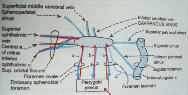
- Cavernous sinus thrombosis
3. Bacteremia – detectable levels of bacteria in blood
4. Septicemia – Increased bacteria + toxins in blood
5. Septic shock – Septicemia, G-ve bacteria, inadequate perfusion of tissues
In summary:
Untreated dental infections can lead to:
A) Local:
- Periapical abscess/granuloma
- Root resorption
- Cellulitis eg. Ludwig’s angina
- Osteomyelitis
- Osteosclerosis
- Trismus
B) Systemic (ascending):
- Cavernous sinus thrombosis
- Meningitis
- Extrusion of orbit
C) Systemic (descending):
- Mediastinitis
- Bacteremia
- Septicemia
- Septic shock
- Necrotising fasciitis

2 thoughts on “Oral Infection – Acute and Chronic”
Comments are closed.