Arteries

Scrotum
Coverings:
- Skin (dark, rugae, thin)
- Superficial fascia (no fat)
- Darto’s muscle
NB: When cold, Darto’s muscle contracts skin, scrotum wrinkles and therefore reduces surface area for heat loss
Scrotum divided internally by Darto’s fascia into left and right, externally seen as scrotal raphe
Location: Inferior and posterior to penis
Contents: Testis, epididymis, spermatic cord
Blood supply: Anterior scrotal, posterior scrotal, cremasteric
Venous: Scrotal veins drain into external pudendal veins
Nerves:
- Genitofemoral nerve – genital branch
- Ilioinguinal nerve – anterior scrotal nerve
- Pudendal nerve – posterior scrotal nerve
- Posterior femoral cutaneous – perineal branch
Lymphatics: Superficial inguinal nodes
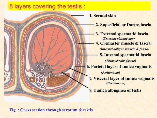
Testis
Coverings:
Blood supply: Testicular, scrotal, deferential, cremasteric
Venous: Right testicular vein drains into IVC, left drains into left renal vein
Nerves: Testicular plexus
Lymphatics: Lumbar nodes
Clinicals:
- Orchitis – inflammation of testis
- Hydrocele – excess fluid in TV
- Hematocele – blood in TV
- Varicocele – venous plexus dilated
- Spermatocele – collection of fluid in epididymis
- Vasectomy – vas deferens ligated and cut
- Distention of scrotum – indirect hernia
Spermatic cord
Forms at deep inguinal ring, enters scrotum via superficial inguinal ring, ends at posterior border of testis
Coverings:
- External spermatic fascia (aponeurosis of external oblique)
- Cremasteric muscle and fascia (internal oblique)
- Internal spermatic fascia (transversalis fascia)
Contents:
- Vas deferens
- Lymph vessels
- Testicular artery
- Cremasteric vessels
- Deferential artery
- Genital nerve
Clinicals:
- Hydrocele of cord
- Torsion of spermatic cord – Surgical emergency, twists on itself, occludes testicular artery and venous drainage leading to necrosis
- Cremasteric reflex

Penis

Coverings:
- Skin – thin, dark, prepuce covers glans
- Deep fascia of penis – continuation of perineal fascia
- Tunica albuginea
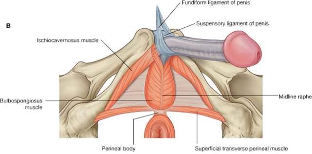
Support:
- Suspensory ligament – connects erectile bodies to pubic symphysis
- Fundiform ligament
Blood supply: Dorsal, deep and bulb of penis arteries
Venous:
- Deep dorsal vein – drains to prostatic venous plexus
- Superficial dorsal vein – drains to superficial external pudendal vein
Nerves:
- Paired dorsal nerve of penis (pudendal nerve) – sensory and sympathetic
- Cavernous nerves (prostatic nerve plexus) – parasympathetic, responsible for the vascular changes which cause erection
Lymphatics: Deep inguinal nodes (glans penis), superficial inguinal nodes
Clinicals:
- Hypospadias – Born with urethra opening on ventral aspect
- Circumcision – surgical excision of prepuce, glans exposed
- Impotence – inability to achieve erection
- Erectile dysfunction – inability to maintain erection
- Priapism – Persistent erection, blood trapped in erectile tissue, can lead to scarring or erectile dysfunction
Prostate gland
Position: Surrounds prostatic urethra, inferior to bladder neck
Relations:
- Anterior – Pubic symphysis
- Posterior – Ampulla of rectum
- Superior – Neck of bladder
- Inferior – External urethral sphincter
- Inferolateral – levator ani muscles
Blood supply: Prostatic artery (from internal iliac), middle rectal, internal pudendal
Venous: Prostatic venous plexus – drains to internal iliac veins
Nerves: Inferior hypogastric plexus
Lymphatics: Internal iliac, sacral nodes
Clinicals:
- Benign prostatic hyperplasia – enlargement of prostate with no malignancy, urinary frequency increases as it compresses bladder and urethra
- Cancer – spread via blood and lymph to IVC, vertebral column and pelvis
Seminal vesicle
Vas deferens combines with seminal vesicle duct to form ejaculatory duct which drains into prostatic urethra
Position: Between bladder fundus and rectum/rectovesical pouch
Relations:
- Anterior – Bladder fundus, ureter
- Posterior – Rectum
- Inferior – Prostate and ejaculatory duct
- Medial – Vas deferens
- Lateral – Prostatic venous plexus
Blood supply: Inferior vesicle, middle rectal, internal pudendal
Nerves: Inferior hypogastric plexus
Lymphatics: External and internal iliac lymph nodes
Clinicals: Seminal gland abscess – may rupture, pus enters peritoneal cavity
Vas deferens
Continuation of epididymis in spermatic cord
Course:
- From tail of epididymis
- Ascends posterior to testis
- Through spermatic cord
- Penetrate abdominal wall via inguinal canal
- Crosses external iliac vessels
- Turns medial between bladder and urethra
- Joins duct of seminal vesicle to form ejaculatory duct
Blood supply: Deferential artery
Venous: Testicular vein, prostate venous plexus
Lymphatics: External iliac
Clinicals: Vasectomy – male sterilization
These are summarized notes from various sources, mainly TeachMeAnatomy and Wikipedia
