Category Archives: Embryology

Development of external genital organs

Indifferent stage

This development occurs between gestational weeks 8 and 12.

  • 2 mesenchymal cloacal folds form on either side of cloacal membrane
  • The 2 cloacal folds fuse infront of the cloacal membrane to form cloacal eminence
  1. Cloacal eminence enlarges – forms genital tubercle
  2. Cloacal membrane divides into urogenital membrane and anal membrane
  3. Cloacal fold divides into genital fold and anal fold
  • Another pair of elevation forms on either side of genital fold – genital swelling
Image result for cloacal folds
development of external genital organs

In females:

  • Genital tubercle – clitoris
  • Genital fold – labia minora
  • Genital swelling – 2 labia majoras

In males:

  • Genital tubercle – elongates to form phallus
  • Genital fold – elongates to form urethral groove which forms urethral plate and then urethral canal
  • Genital swelling – 2 scrotal swellings, fuse – scrotum

Congenital anomalies:

  1. Absence of penis/clitoris – no genital tubercle develops
  2. Small penis – underdeveloped genital tubercle
  3. large clitoris – overdeveloped genital tubercle
  4. Divided scrotum – failure of genital swelling to fuse
  5. False hermaphrodite – gonads are of one sex, external genitalia are of opposite sex

Development of genital system

Indifferent gonads

  • A genital ridge forms from intermediate mesoderm (medial to mesonephros) – forms stroma of gonads
  • Primitive sex cords form from mesodermal coelomic epithelium covering the genital ridge
  • Primordial germ cells (endodermal) develop in wall of yolk sac ⇒ pass through dorsal mesentery ⇒ lie inbetween primitive sex cords
  • Gonads don’t acquire male or female characteristics until week 7
Image result for primitive sex cords and yolk sac

Testis

  • Primitive sex cords branch and anastomose to from testis cordis
  • Primordial germ cells incorporate in the testis cordis
  • Testis cordis lose connection with the surface epithelium – form seminiferous tubules
  • Straight ends of seminiferous tubules anastomose at hilum of testis and form rete testis
  • Rete testis connect to mesonephric duct via 8-12 mesonephric tubules – forms head of epididymis
  • Surface epithelium disappears, testis surrounded by thick fibrous capsule – tunica albuginea
Image result for primitive sex cords and yolk sac

Descent of testis:

  • Testis develop on posterior abdominal wall behind peritoneum
  • A gubernacular cord extends from lower pole of testis to scrotal pouch
  • Gubernacular cord shortens due to chorionic gonadotropins and increased intraabdominal pressure
  • Goes through inguinal canal
  • Remnant of gubernaculum disappears
  • An evagination of peritoneal cavity called vaginal process enters scrotum
  • Forms serous cavity for testis called tunica vaginalis
  • Proximal part of vaginal process obliterated
descent of testis

Anomalies of testis:

  1. Cryptorchidism – undescended testis
  2. Maldescended testis – lying somewhere over the normal line of descent
  3. Ectopic testis – outside that line
  4. Congenital inguinal hernia – failure of obliteration of proximal vaginal process

Male genital ducts

1. Mesonephric tubules:

  • Upper – degenerate, form appendix of epididymis
  • Middle – 6-12 form head of epididymis and are connected to rete testis
  • Lower – degenerate, form paradidymis

2. Mesonephric duct:

  • Body and tail of epididymis
  • Vas deferens
  • Seminal vesicle
  • Ejaculatory duct

3. Mullerian duct (notes in female):

Degenerates completely except the upper end – forms appendix of testis


Ovaries

  • Primitive sex cords break into clusters of cells – form primary medullary cords
  • Which is replaced by vascular stroma to form – medulla of ovary
  • Coelomic epithelium proliferates again – forms 2nd generation of ovary (sex) cords
  • Which will divide into clusters of cells – follicular cells of primary follicle
  • Primordial germ cells incorporate into primary follicles – form oogonia
  • Primitive cortex becomes secondary cortex containing primary follicles
  • Medulla is just vascular stroma, no follicles
Image result for development of ovaries

Descent of ovaries:

  • Ovary develops in posterior abdominal wall
  • Gubernacular cord from lower pole of ovary to labia majora
  • Pulls ovary to its level on pelvis
  • Uterus develops, gubernaculum divides into 2 parts:
  1. Ovarian ligament – ovary to uterus
  2. Round ligament – uterus to labia majora (goes through inguinal canal)

Congenital anomalies of ovaries:

  1. Congenital absence – Turner’s syndrome
  2. True hermaphroditism – gonads of both sex present
  3. Imperfect descent – in inguinal canal
  4. Vagina agenesis

Female genital ducts

  • 2 mullerian (paramesonephric) ducts arise from coelomic epithelium, lateral to mesonephric ducts
  • Grow caudally, curve medially infront of mesonephric duct, meet each other and grow caudally
development of genital system - female genital ducts
  • The 2 ducts fuse to form uterovaginal canal
  • The lower tip of uterovaginal canal grows downwards and protrudes posterior wall of urogenital sinus
  • 2/3 of mullerian ducts form oviducts
  • Uterovaginal canal forms uterus and upper 4/5 vagina
  • Where the 2 ducts unite, forms fundus of uterus
  • Lower 1/5 vagina forms from definitive urogenital sinus

Union between upper 4/5 and lower 1/5 vagina is demarcated by hymen

Mesonephric tubules and mesonephric duct degenerates

Congenital anomalies:

  1. Double uterus, double vagina – complete failure of fusion
  2. Double uterus, single vagina – partial failure of fusion
  3. Agenesis of uterus – failure of both mullerian ducts to develop
  4. Rudimentary horn – failure of one mullerian ducts to develop, therefore one fallopian tube, and half body of uterus connected to rudimentary horn
  5. Atresia of cervix/vagina
  6. Imperforate hymen – cells between junction fail to degenerate
  7. Remnants of mesonephric tubules – enlarge and form cysts
  8. Remnants of mesonephric duct – Gartner’s duct
  9. Infantile uterus – small uterus, large cervix
Image result for Gartner's duct

Development of urinary system

Development of kidney, urinary bladder and urethra


Kidney

  • Intermediate mesoderm: kidneys, ureters and trigone of urinary bladder
  • Endoderm: rest of urinary bladder, urethra

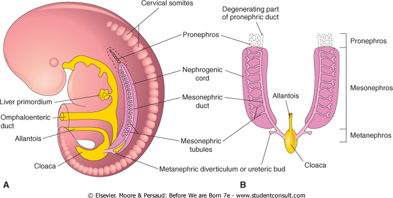
3 kidneys develop: Pronephros, mesonephros and metanephros

development of kidneys - mesonephros
Image result for pronephros, mesonephros, metanephros
development of kidney

(I) Pronephros:

  • Segmented cervical intermediate mesoderm
  • 7-10 excretory tubules called pronephric tubules form – degenarate by end of 4th week
  • Collecting duct called pronephric duct forms and opens down into cloaca – pronephric duct persists to form mesonephric duct

(II) Mesonephros:

  • Segments of thoracic and upper lumbar region of intermediate mesoderm
  • Each segment forms 2-3 ‘S’ shaped mesonephric tubules
  • Lateral ends open in mesonephric duct and medial end invaginated by glomerulus

In males:

  • Mesonephric tubules: upper degenerate, rest form efferent ducts of testis, head of epididymis, paradidymis
  • Mesonephric duct: body and tail of epididymis, vas deferens, ejaculatory duct, seminal vesicle, ureteric bud and trigone of urinary bladder

In females:

  • Mesonephric tubules: degenerate
  • Mesonephric duct: ureteric bud and trigone of urinary bladder

(III) Metanephros: (kidneys)

(A) Development of collecting duct and ureter:

  • Ureteric bud develops from mesonephric duct
  • Ureteric bud grows cranially, and penetrates metanephric cap
  • Upper end of ureteric bud enlarges ⇒ forms pelvis which divides into ⇒ 2-3 major calyces ⇒ where each divides into minor calyces ⇒ then collecting tubules ⇒ which join to nephrons 
Image result for development of urinary bladder
Image result for development of kidney

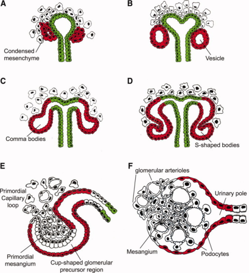
(B) Development of nephrons:

  • Caudal part of intermediate mesoderm forms a metanephric cap
  • Which divides into renal vesicles
  • Each renal vesicle surrounds the free end of a collecting tubule and forms a ‘S’ shaped nephron
  • One end of nephron invaginated by glomerulus – Bowman’s capsule
  • Other end joins collecting duct
  • Each nephron elongates – forms proximal and distal convolutes tubules and loop of Henle
Image result for renal vesicles
development of nephrons

Further growth of kidney:

  1. Lobulated grooves disappear – forms smooth surface
  2. Ascends from pelvic region to adult level
  3. Recieves blood supply from median sacral, common iliac, lower abdominal aorta. Then only from aorta
  4. At first, hilum directed forwards, rotates 90 degrees so hilum becomes medial

Congenital anomalies of kidney:

  1. Renal agenesis
  2. Renal hypogenesis – small size
  3. Congenital polycystic kidney – failure of fusion between nephrons and collecting tubules. Urine collects in nephrons, dilates and forms cysts, nephrons destroyed
  4. Pelvic kidney – failure of ascent
  5. Horseshoe shaped kidney – fusion of both kidneys, ureters kinked, this causes urinary stasis and so infection
  6. Additional branches of aorta supplying kidney – cross infront of ureter and compress it – urinary stasis
  7. Double ureter – 2 ureteric buds/ early splitting of ureteric bud. More liable to infection and stone formation
Image result for Congenital polycystic kidney

Urinary bladder and urethra

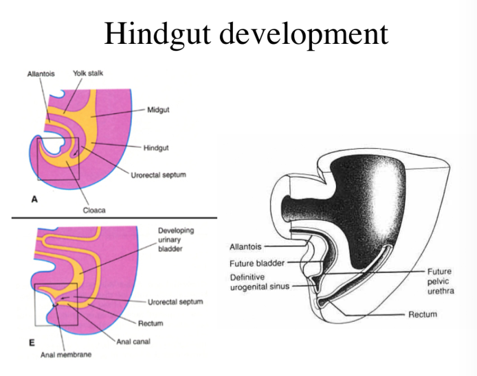
A constriction appears in primitive urogenital sinus at site of entrance of mesonephric duct

Divides into:

  1. Upper part – Vesico-urethral canal
  2. Lower part – Definitive urogenital sinus

(I) In males

(A) Urinary bladder:

  • From vesico-urethral canal
  • Trigone from absorbed common stem of mesonephric duct and ureter. 

Differential growth of posterior bladder wall, therefore ureter moves upwards (posterior superior angle)

(B) Seminal vesicle:

Develops as a diverticulum from vas deferens. Part distal to it becomes ejaculatory duct.

(C) Urethra:

1. Prostatic urethra:

  • Upper 1/2 – vesicourethral canal
  • Lower 1/2 – Definitive urogenital sinus

2. Membranous urethra – Definitive urogenital sinus

3. Penile urethra – Definitive urogenital sinus forms a urethral plate that extends on the under surface of phallus (primitive penis) and is surrounded by 2 urethral folds – unite from back and front around urethral plate to form penile urethra. Lined by endoderm, terminal glandular part lined by ectoderm.

(D) Prostate gland:

  • Develops from 15 to 20 buds from prostatic urethra
  • Canalized to form alveoli and ducts
  • Connective tissue and capsule from surrounding mesoderm

(II) In females

  1. Vesicourethral canal – urinary bladder and urethra
  2. Definitive urogenital sinus – lower 1/5 vagina and vestibule

Congenital anomalies:

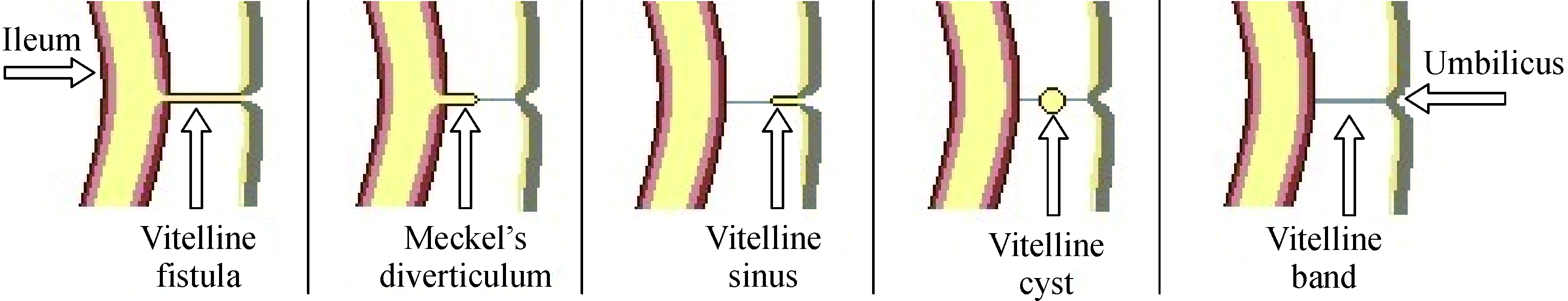
  1. Urachal fistula – unobliterated urachus. Urine drips from umbilicus
  2. Urachal cyst – Incomplete obliteration
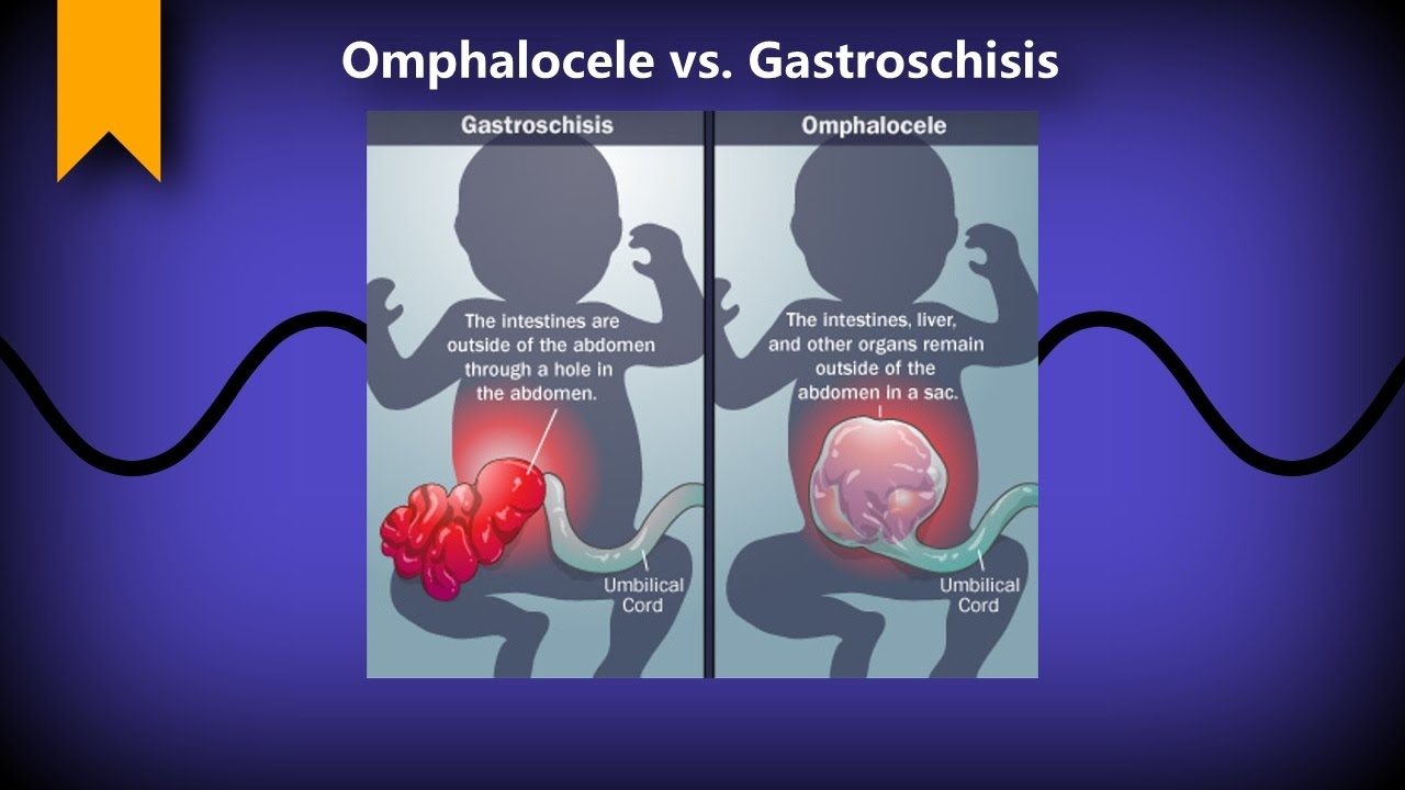
  3. Bladder extrophy – urinary bladder opens into anterior abdominal wall
  4. Hypospadius – external urethral meatus opens on under surface of penis
  5. Epispadius –  external urethral meatus opens on upper surface of penis

Development of midgut and hindgut

Midgut

Origin: Endodermal

development of midgut and hindgut
  1. Cranial limb – forms jejunum and ileum
  2. Caudal limb – forms ascending colon and 2/3 of transverse colon
  3. Cecal swelling – forms cecum, appendix and part of ascending colon
  • Intestinal loop elongates rapidly and leaves the small abdominal cavity and enters umbilical cord – physiological umbilical hernia (6th to 10th week)
  • The elongating loop rotates 270 degrees anticlockwise around axis of superior mesenteric artery (seen in the diagram)
  • Therefore upper part of small intestine lies behind colon
  • 10th week, abdominal cavity enlarges and :
  1. Jejunum reenters to left side
  2. Ileum reenters to right side
  3. Cecal swelling reenters below liver
  • Cecal swelling elongates downwards to right iliac fossa – forms right colic flexure and ascending colon
  • Vitellointestinal duct obliterated

Congenital anomalies:

  1. Remnant of vitelline duct:
  • Meckel’s diverticulum – proximal part near ileum remains patent
  • Vitelline fistula – whole vitelline duct remains open
  • Vitelline cyst – Middle part remains open
Image result for vitelline cyst

2. Omphalocele/ Congenital umbilical hernia – Failure of reduction of physiological hernia due to defect in abdominal wall muscles development

3. Gastroschisis

Image result for omphalocele vs gastroschisis

4. Atresia/ stenosis of any part of primitive intestinal loop – bowel obstruction

5. Abdominal rotation of intestinal loop  – 90 degrees only or clockwise rotation


Hindgut

Origin: Endodermal

Derivatives: Left 1/3 transverse colon, descending colon, sigmoid colon, rectum, upper 1/2 anal canal (Lower 1/2 anal canal, proctodeum – ectodermal)

  • Lower end of hindgut dilates to form cloaca
  • And then continues as allantois to umbilicus
  • Below cloaca is cloacal membrane
  • Which is bilaminar: outer ectoderm, inner endoderm
  • Between hindgut and allantois is a urorectal septum, which grows caudally and divides the cloaca and cloacal membrane  into:
  1. Primitive urogenital sinus (ventrally) – Urogenital membrane
  2. Rectoanal canal (dorsally) – anal membrane
  • Opposite rectoanal canal, ectodermal depression called proctodeum forms
  • Anal membrane ruptures:
  1. Proctodeum – lower 1/2 anal canal
  2. Rectoanal canal – rectum and upper 1/2 anal canal
development of hindgut
Hindgut development

Congenital anomalies:

  1. Imperforate anus – anal membrane fails to rupture
  2. Atresia of rectum – Proctodeum fails to develop
  3. Stenosis of rectum – incomplete canalization
  4. Recto – vaginal fistula, recto – urinary fistula, recto – urethral fistula – incomplete division of cloaca
  5. Anal atresia, anal stenosis
  6. Ectopic anus

Development of septum transversum and diaphragm

Septum transversum

It is a thick mass of mesoderm which partially separates thoracic cavity and abdominal cavity.

  • Forms in neck by fusion of 3, 4, 5 cervical myotomes
  • Motor nerve is phrenic nerve
  • Embryonic disc folds and heart descends, therefore septum transversum is pushed caudally and pulls the phrenic nerve with it

Derivatives:

  1. Superior layer – formation of fibrous pericardium
  2. middle layer – diaphragm muscle, central tendon, diaphragmatic pleura and peritoneum
  3. Inferior layer – fibrous capsule and connective tissue of liver, ventral mesentery of the gut


Diaphragm

Origin: Mesoderm

Diaphragm develops from:

  1. Septum transversum – Central tendon, sternal and costal parts of diaphragm
  2. 2 pleuro-peritoneal membranes – 2 mesodermal folds that project inwards from body wall. Close pleuro-peritoneal canals. Forms dorsilateral part of diaphragm
  3. Mesoderm from chest wall – marginal part of diaphragm
  4. Mesentery of esophagus – Posterior medial part and crura of diaphragm
  5. Mesoderm around aorta – lumbar part of diaphragm


Image result for development of diaphragm

Congenital anomalies:

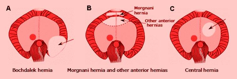
  1. Parasternal hernia of Morgagni – failure to develop a small part of diaphragm between sternal and costal part
  2. Esophageal hernia – Protrusion of stomach in thorax
  3. Congenital diaphragmatic hernia of Bochdalek – failure of pleuro-peritoneal membranes to close the pleuro-peritoneal canals. Abdominal vicera enter pleural cavity, compress heart and lungs

Image result for Parasternal hernia of Morgagni

Development of pituitary gland and SNS

Development of pituitary gland and sympathetic nervous system

Pituitary gland

Anterior lobe (adenohypophysis)

  • Ectoderm – roof of stomodeum 
  • Forms diverticulum called Rathke’s pouch, which grows upwards from stomodeum infront of buccopharyngeal membrane
  • End of 3rd month, loses connection with stomodeum and differentiates into; Pars distalis, Pars tuberalis and Pars intermedia
  • Lumen gets obliterated

Posterior lobe (neurohypophysis)

  • Neural ectoderm – floor of diencephalon
  • Forms diverticulum called Infundibulum, which grows downwards
  • Differentiates into; Pars nervosa and pituitary stalk

Development of pituitary gland

Congenital anomalies:

  1. Agenesis
  2. Absence of anterior lobe – failure of developing Rathke’s pouch. Leads to maldeveloped thyroid gland, suprarenal gland and testes
  3. Pharyngeal hypophysis – remnant of Rathke’s pouch remains attatched to pharyngeal wall

Sympathetic nervous system

  • Neural crest cells in thoracic region
  • Migrate to each side of the spinal cord, behind dorsal aorta
  • Form a bilateral chain of segmentally arranged sympathetic ganglia which are connected by longitudinal nerve fibers
  • Neuroblasts migrate from thorax region to cervical and lumbosacral regions, therefore extending the sympathetic chains

In cervical region, ganglia fuse to form superior, middle and inferior cervical ganglia

Some neuroblasts migrate infront of aorta to form celiac, superior mesenteric and inferior mesenteric ganglia

Some neuroblasts migrate to heart, lungs or GIT to form sympathetic organ plexuses

Some migrate to form medulla of suprarenal glands

Development of spleen and suprarenal glands

Spleen

Origin: Mesodermal cells in dorsal mesogastrium

  • These cells will form stroma and capsule
  • Hematopoietic cells infiltrate spleen
  • Dorsal mesogastrium forms: gastrosplenic ligament and splenorenal ligament

NB: Hematopoietic function lost with embryo development. Lymphoid precursor cells migrate into developing organ

development of spleen

Congenital anomalies:

  1. Accessory spleen
  2. Wandering spleen – lacks one ligament or both
  3. Polysplenia/ Chaudhrey’s disease – multiple small accessory spleens

Suprarenal glands

  • Cortex – Mesodermal cells of intraembryonic coelomic epithelium
  • On either side of mesentery of gut, proliferates to form fetal cortex
  • Medulla – Sympatho – chromaffin cells from neural crest cells (ectoderm)
  • Migrate to enter medial aspect of fetal cortex
  • Second layer of cells develop from coelomic mesothelium and surrounds fetal cortex to form permanent cortex
  • Fetal cortex regresses and disappears after 3rd year of birth
  • Permanent cortex differentiates into 3 zones: zona glomerulosa, zona fasiculata and zona reticularis. Complete histological differentiation attained at puberty.
Image result for development of suprarenal gland

Congenital anomalies:

  1. Agenesis
  2. Ectopic suprarenal gland – below capsule of kidney
  3. Accessory cortical tissue – found on posterior abdominal wall and pelvis
  4. Adrenogenital syndrome – hypertrophy of suprarenal cortex and over production of androgens. Results of pseudohermaphroditism in females and premature enlargement of external genitalia in males

Development of liver and gall bladder

Origin: Endoderm of foregut

  • Liver bud develops from lower end of foregut
  • Liver bud divides into two parts:
  1. Smaller part: Pars cystica ⇒ forms gall bladder
  2. Large cranial part: Pars hepatica
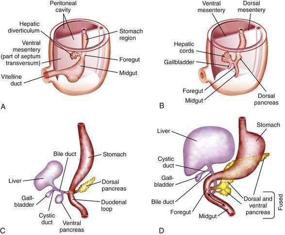
  • Pars hepatica invades septum transversum and divides into right and left branches (right and left hepatic ducts) which branch more to form columns of hepatic cells
  • Columns of hepatic cells meet vitelline veins and break them into hepatic sinusoids
  • Mesoderm of septum transversum forms fibrous tissue stroma and capsule of liver
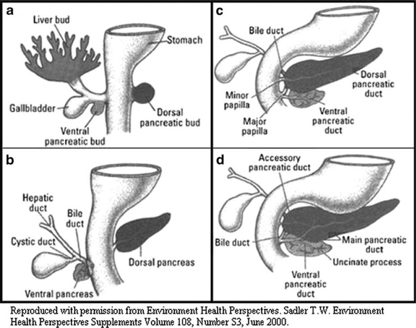
  • Original stalk of liver bud elongates – forms common bile duct
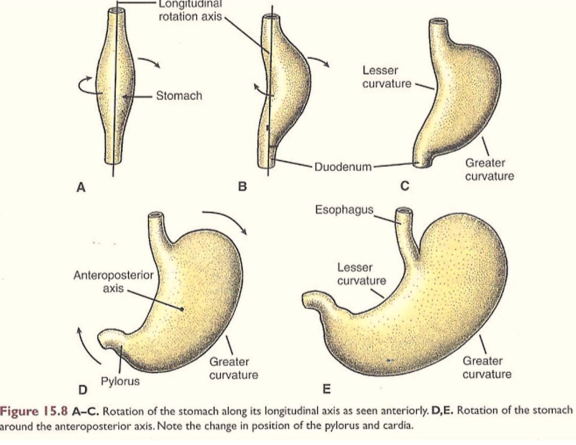
  • Due to rotation of stomach, common bile duct opens in posterior medial part of 2nd duodenum

Ligaments of liver:

  • Mesoderm of septum transversum between liver and anterior abdominal wall forms falciform ligament. Umbilical vein lies on inferior free margin of falciform ligament
  • Mesoderm of septum transversum between liver and stomach forms lesser omentum

Liver separates from septum transversum except “bare area” of liver

Rest of septum transversum forms part of diaphragm

Image result for Development of liver and gall bladder

Congenital anomalies:

  1. Atresia of common bile duct
  2. Partial or complete duplication of gall bladder
  3. Congenital absence of portal vein
  4. Accessory hepatic duct

Development of stomach, duodenum and pancreas

Stomach

Origin: Endodermal

  • Lower part of foregut, swelling appears
  • Anterior border connected to anterior abdominal wall by ventral mesogastrium
  • Posterior border connected to posterior abdominal wall by dorsal mesogastrium
  • Posterior border grows more – forms greater curvature of stomach
  • Anterior border forms lesser curvature
  • Stomach then rotates 90 degrees clockwise (due to growth of liver)
  • So dorsal mesogastrium forms lesser sac
  • Liver develops in ventral mesentery – mesentery becomes lesser omentum
  • Spleen develops in dorsal mesentery – mesentery becomes gastrosplenic ligament

Clinicals: Congenital hypertrophy of pyloric sphincter – narrows pyloric canal. More common in males.

NB: Development of stomach and rotation causes right vagus nerve to become posterior vagus trunk. Left vagus nerve becomes anterior vagus trunk

Image result for development of stomach

Duodenum

Origin: Endodermal

  • Distal foregut and proximal midgut
  • Form a “C” shaped loop projecting ventrally
  • Due to stomachs rotation 90 degrees clockwise
  • Duodenal loop carried dorsally and to the right, becomes adherent to posterior abdominal wall
  • Mesentery of duodenum disappears except in the 1st and 4th part (ligament of trietz)
  • The lumen is temporarily obstructed and canalized again

Congenital anomalies:

  1. Incomplete canalization/ atresia – green vomit, no bowel movement
  2. Incomplete fixation to posterior abdominal wall – sites of internal hernia
Image result for development of duodenum


Pancreas

Origin: 2 endodermal buds form:

  1. Endoderm of dorsal wall of duodenum – dorsal pancreatic bud
  2. Endoderm of ventral wall of duodenum from hepatic bud stem – ventral pancreatic bud
  • Dorsal pancreatic bud arises slightly above liver bud, extends dorsally and upwards into mesoduodenum
  • Ventral pancreatic bud migrates dorsally to lie below and behind dorsal bud
  • The 2 buds fuse together (ventral bud – head and uncinate process)
  • Ducts of the 2 buds join – main pancreatic duct
  • Islets of Langerhans appear – insulin secretion begins in 5th month

Congenital anomalies:

  1. Annular pancreas – develops as a ring around 2nd part duodenum
  2. Ectopic pancreatic tissue – eg. wall of duodenum, jejunum, ileum or stomach
development of pancreas

Development of eyes and ears

Development of Eyes

Development of eyes

(I) Retina and optic nerve

  • Optic vesicle of prosencephalon extends laterally
  • Contacts surface ectoderm
  • Optic vesicle invaginated to form optic cup:
  1. Outer layer – forms pigmented layer of retina
  2. Inner layer anterior 1/5 – iris and ciliary body
  3. Inner layer posterior 4/5 – All layers of retina except pigmented layer
  • Mouth of optic cup forms pupil
  • Optic stalk invaginated by central artery of retina – gets enclosed in optic stalk – forms optic nerve

(II) Lens

  • Surface ectoderm thickens – forms lens placode
  • Lens placode invaginated at optic cup mouth – forms lens vesicle
  • Lens vesicle separates from surface ectoderm
  • Lies in optic cup mouth
  • Cells of posterior layer of lens vesicle elongate – forms lens fibers

(III) Choroid and sclera – Forms from mesenchyme around optic vesicle


Development of Ear

Develops from pharyngeal arches (link)

Develops around 4th and 5th week of development, while the middle ear ossicles form around 6 weeks of development.

Muscles of middle ear – tensor tympani (1st arch) and stapedius muscle (2nd arch)