Development of kidney, urinary bladder and urethra
Kidney
- Intermediate mesoderm: kidneys, ureters and trigone of urinary bladder
- Endoderm: rest of urinary bladder, urethra
3 kidneys develop: Pronephros, mesonephros and metanephros

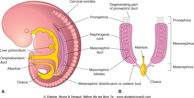
(I) Pronephros:
- Segmented cervical intermediate mesoderm
- 7-10 excretory tubules called pronephric tubules form – degenarate by end of 4th week
- Collecting duct called pronephric duct forms and opens down into cloaca – pronephric duct persists to form mesonephric duct
(II) Mesonephros:
- Segments of thoracic and upper lumbar region of intermediate mesoderm
- Each segment forms 2-3 ‘S’ shaped mesonephric tubules
- Lateral ends open in mesonephric duct and medial end invaginated by glomerulus
In males:
- Mesonephric tubules: upper degenerate, rest form efferent ducts of testis, head of epididymis, paradidymis
- Mesonephric duct: body and tail of epididymis, vas deferens, ejaculatory duct, seminal vesicle, ureteric bud and trigone of urinary bladder
In females:
- Mesonephric tubules: degenerate
- Mesonephric duct: ureteric bud and trigone of urinary bladder
(III) Metanephros: (kidneys)
(A) Development of collecting duct and ureter:
- Ureteric bud develops from mesonephric duct
- Ureteric bud grows cranially, and penetrates metanephric cap
- Upper end of ureteric bud enlarges ⇒ forms pelvis which divides into ⇒ 2-3 major calyces ⇒ where each divides into minor calyces ⇒ then collecting tubules ⇒ which join to nephrons


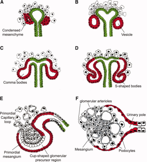
(B) Development of nephrons:
- Caudal part of intermediate mesoderm forms a metanephric cap
- Which divides into renal vesicles
- Each renal vesicle surrounds the free end of a collecting tubule and forms a ‘S’ shaped nephron
- One end of nephron invaginated by glomerulus – Bowman’s capsule
- Other end joins collecting duct
- Each nephron elongates – forms proximal and distal convolutes tubules and loop of Henle
Further growth of kidney:
- Lobulated grooves disappear – forms smooth surface
- Ascends from pelvic region to adult level
- Recieves blood supply from median sacral, common iliac, lower abdominal aorta. Then only from aorta
- At first, hilum directed forwards, rotates 90 degrees so hilum becomes medial
Congenital anomalies of kidney:
- Renal agenesis
- Renal hypogenesis – small size
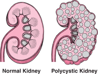
- Congenital polycystic kidney – failure of fusion between nephrons and collecting tubules. Urine collects in nephrons, dilates and forms cysts, nephrons destroyed
- Pelvic kidney – failure of ascent
- Horseshoe shaped kidney – fusion of both kidneys, ureters kinked, this causes urinary stasis and so infection
- Additional branches of aorta supplying kidney – cross infront of ureter and compress it – urinary stasis
- Double ureter – 2 ureteric buds/ early splitting of ureteric bud. More liable to infection and stone formation
Urinary bladder and urethra
A constriction appears in primitive urogenital sinus at site of entrance of mesonephric duct
Divides into:
- Upper part – Vesico-urethral canal
- Lower part – Definitive urogenital sinus
(I) In males
(A) Urinary bladder:
- From vesico-urethral canal
- Trigone from absorbed common stem of mesonephric duct and ureter.
Differential growth of posterior bladder wall, therefore ureter moves upwards (posterior superior angle)
(B) Seminal vesicle:
Develops as a diverticulum from vas deferens. Part distal to it becomes ejaculatory duct.
(C) Urethra:
1. Prostatic urethra:
- Upper 1/2 – vesicourethral canal
- Lower 1/2 – Definitive urogenital sinus
2. Membranous urethra – Definitive urogenital sinus
3. Penile urethra – Definitive urogenital sinus forms a urethral plate that extends on the under surface of phallus (primitive penis) and is surrounded by 2 urethral folds – unite from back and front around urethral plate to form penile urethra. Lined by endoderm, terminal glandular part lined by ectoderm.
(D) Prostate gland:
- Develops from 15 to 20 buds from prostatic urethra
- Canalized to form alveoli and ducts
- Connective tissue and capsule from surrounding mesoderm
(II) In females
- Vesicourethral canal – urinary bladder and urethra
- Definitive urogenital sinus – lower 1/5 vagina and vestibule
Congenital anomalies:
- Urachal fistula – unobliterated urachus. Urine drips from umbilicus
- Urachal cyst – Incomplete obliteration
- Bladder extrophy – urinary bladder opens into anterior abdominal wall
- Hypospadius – external urethral meatus opens on under surface of penis
- Epispadius – external urethral meatus opens on upper surface of penis
