Indifferent gonads
- A genital ridge forms from intermediate mesoderm (medial to mesonephros) – forms stroma of gonads
- Primitive sex cords form from mesodermal coelomic epithelium covering the genital ridge
- Primordial germ cells (endodermal) develop in wall of yolk sac ⇒ pass through dorsal mesentery ⇒ lie inbetween primitive sex cords
- Gonads don’t acquire male or female characteristics until week 7

Testis
- Primitive sex cords branch and anastomose to from testis cordis
- Primordial germ cells incorporate in the testis cordis
- Testis cordis lose connection with the surface epithelium – form seminiferous tubules
- Straight ends of seminiferous tubules anastomose at hilum of testis and form rete testis
- Rete testis connect to mesonephric duct via 8-12 mesonephric tubules – forms head of epididymis
- Surface epithelium disappears, testis surrounded by thick fibrous capsule – tunica albuginea

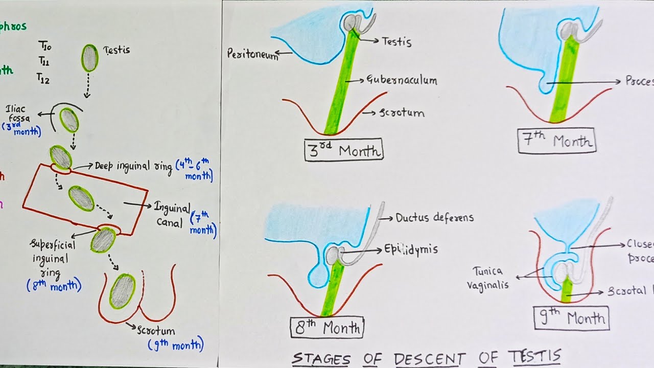
Descent of testis:
- Testis develop on posterior abdominal wall behind peritoneum
- A gubernacular cord extends from lower pole of testis to scrotal pouch
- Gubernacular cord shortens due to chorionic gonadotropins and increased intraabdominal pressure
- Goes through inguinal canal
- Remnant of gubernaculum disappears
- An evagination of peritoneal cavity called vaginal process enters scrotum
- Forms serous cavity for testis called tunica vaginalis
- Proximal part of vaginal process obliterated
Anomalies of testis:
- Cryptorchidism – undescended testis
- Maldescended testis – lying somewhere over the normal line of descent
- Ectopic testis – outside that line
- Congenital inguinal hernia – failure of obliteration of proximal vaginal process
Male genital ducts
1. Mesonephric tubules:
- Upper – degenerate, form appendix of epididymis
- Middle – 6-12 form head of epididymis and are connected to rete testis
- Lower – degenerate, form paradidymis
2. Mesonephric duct:
- Body and tail of epididymis
- Vas deferens
- Seminal vesicle
- Ejaculatory duct
3. Mullerian duct (notes in female):
Degenerates completely except the upper end – forms appendix of testis
Ovaries
- Primitive sex cords break into clusters of cells – form primary medullary cords
- Which is replaced by vascular stroma to form – medulla of ovary
- Coelomic epithelium proliferates again – forms 2nd generation of ovary (sex) cords
- Which will divide into clusters of cells – follicular cells of primary follicle
- Primordial germ cells incorporate into primary follicles – form oogonia
- Primitive cortex becomes secondary cortex containing primary follicles
- Medulla is just vascular stroma, no follicles

Descent of ovaries:
- Ovary develops in posterior abdominal wall
- Gubernacular cord from lower pole of ovary to labia majora
- Pulls ovary to its level on pelvis
- Uterus develops, gubernaculum divides into 2 parts:
- Ovarian ligament – ovary to uterus
- Round ligament – uterus to labia majora (goes through inguinal canal)
Congenital anomalies of ovaries:
- Congenital absence – Turner’s syndrome
- True hermaphroditism – gonads of both sex present
- Imperfect descent – in inguinal canal
- Vagina agenesis
Female genital ducts
- 2 mullerian (paramesonephric) ducts arise from coelomic epithelium, lateral to mesonephric ducts
- Grow caudally, curve medially infront of mesonephric duct, meet each other and grow caudally

- The 2 ducts fuse to form uterovaginal canal
- The lower tip of uterovaginal canal grows downwards and protrudes posterior wall of urogenital sinus
- 2/3 of mullerian ducts form oviducts
- Uterovaginal canal forms uterus and upper 4/5 vagina
- Where the 2 ducts unite, forms fundus of uterus
- Lower 1/5 vagina forms from definitive urogenital sinus
Union between upper 4/5 and lower 1/5 vagina is demarcated by hymen
Mesonephric tubules and mesonephric duct degenerates
Congenital anomalies:
- Double uterus, double vagina – complete failure of fusion
- Double uterus, single vagina – partial failure of fusion
- Agenesis of uterus – failure of both mullerian ducts to develop
- Rudimentary horn – failure of one mullerian ducts to develop, therefore one fallopian tube, and half body of uterus connected to rudimentary horn
- Atresia of cervix/vagina
- Imperforate hymen – cells between junction fail to degenerate
- Remnants of mesonephric tubules – enlarge and form cysts
- Remnants of mesonephric duct – Gartner’s duct
- Infantile uterus – small uterus, large cervix

