Septum transversum
It is a thick mass of mesoderm which partially separates thoracic cavity and abdominal cavity.
- Forms in neck by fusion of 3, 4, 5 cervical myotomes
- Motor nerve is phrenic nerve
- Embryonic disc folds and heart descends, therefore septum transversum is pushed caudally and pulls the phrenic nerve with it
Derivatives:
- Superior layer – formation of fibrous pericardium
- middle layer – diaphragm muscle, central tendon, diaphragmatic pleura and peritoneum
- Inferior layer – fibrous capsule and connective tissue of liver, ventral mesentery of the gut
Diaphragm
Origin: Mesoderm
Diaphragm develops from:
- Septum transversum – Central tendon, sternal and costal parts of diaphragm
- 2 pleuro-peritoneal membranes – 2 mesodermal folds that project inwards from body wall. Close pleuro-peritoneal canals. Forms dorsilateral part of diaphragm
- Mesoderm from chest wall – marginal part of diaphragm
- Mesentery of esophagus – Posterior medial part and crura of diaphragm
- Mesoderm around aorta – lumbar part of diaphragm

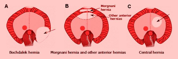
Congenital anomalies:
- Parasternal hernia of Morgagni – failure to develop a small part of diaphragm between sternal and costal part
- Esophageal hernia – Protrusion of stomach in thorax
- Congenital diaphragmatic hernia of Bochdalek – failure of pleuro-peritoneal membranes to close the pleuro-peritoneal canals. Abdominal vicera enter pleural cavity, compress heart and lungs