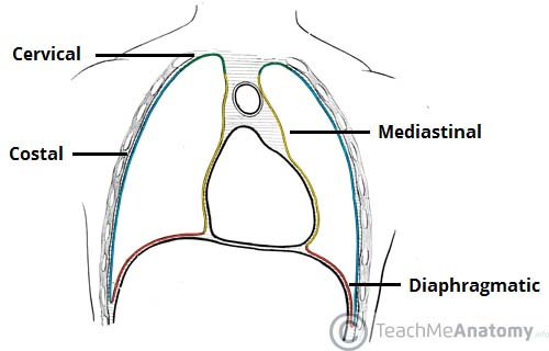
Lung’s pleura
2 pleuras: continuous at hilum
- Parietal pleura – lines inner surface of the thoracic cavity
- Visceral pleura – lines surface of lung
Pleural cavity: contains pleural fluid which lubricates lungs
4 parts of parietal pleura:
Pleural recesses:

Clinicals:
- Drain fluid – insert needle superior to rib
- Pleuritis
- Pancoast tumor – on lung apex, erodes 1st rib
- Pyothorax (pus), hemothorax (blood), pneumothorax (air), chylothorax (lymph) – collect in pleural cavity
Lungs
Lung surfaces: Costal, mediastinal, diaphragmatic
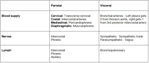
Blood supply: Same as visceral pleura
Nerve: Same as visceral pleura
- Sympathetic trunk: Relax bronchial smooth muscle, vasoconstrict vessels
- Vagus: Contract smooth muscles, vasodilate
Bronchial tree: Trachea ⇒ Right and left bronchus ⇒ Lobar bronchus (3 right, 2 left) ⇒ Segmental bronchus ⇒ Interlobular bronchus ⇒ Terminal bronchiole ⇒ Respiratory bronchiole
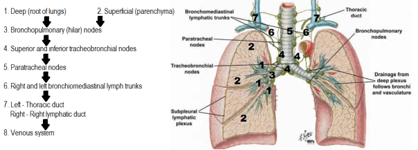
Lymphatics:
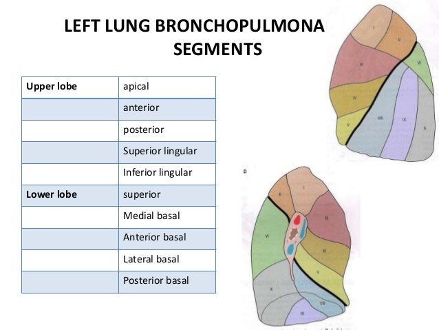
Bronchopulmonary segment:
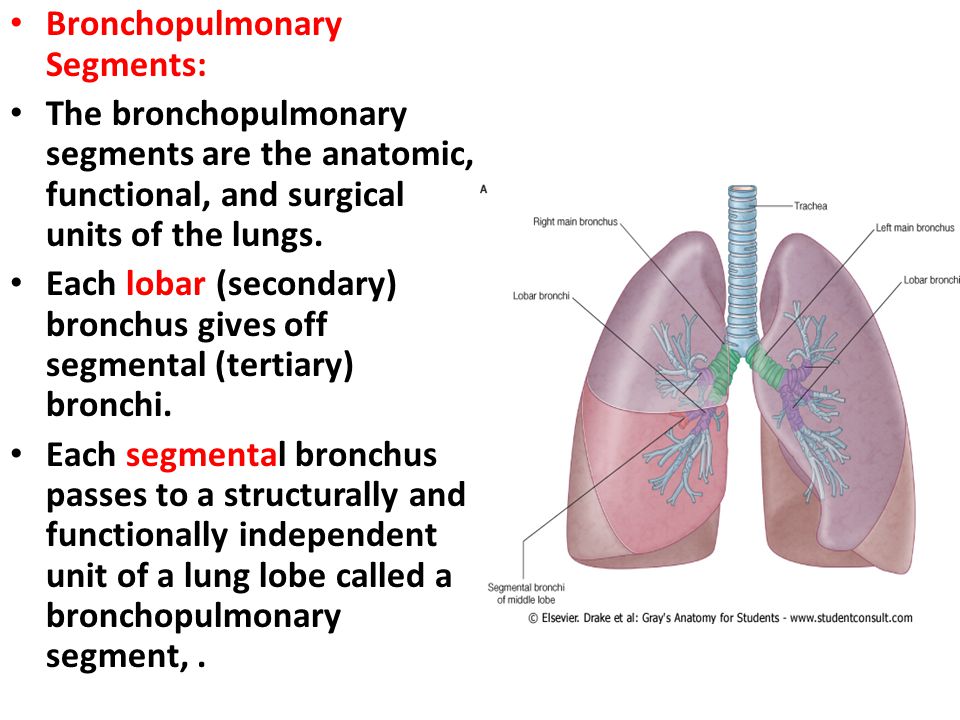
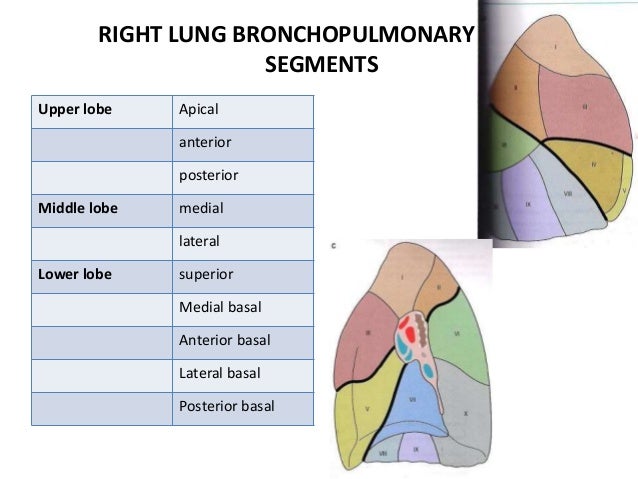
Clinicals: Lung resection – remove specific tumor on segment
Apex of lung – relations:
- Anterior – subclavian artery, scalenus anterior, clavicle
- Posterior – posterior intercostal arteries and veins
- Lateral – 1st rib
- Medial – phrenic nerve, vagus nerve, trachea, esophagus
- Superior – brachial plexus
Right and left lung differences:
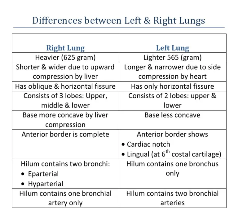
Right – 10 segments, Left – 8 segments
Right and left bronchi:
- Right: Shorter, vertical, 3 divisions, enters lung at T5 level
- Left: Longer, horizontal, 2 divisions, enters lung at T6 level
Right and left hilum:

Right and left lung impressions:

Clinicals:
- Pulmonary embolism – dyspnea, chest pain, cough blood
- Clavicle fractures – damage apex of lungs
- Asthma
- Chronic bronchitis
- Cancer – smoking
- Cystic fibrosis
- Bronchoscopy
- Aspiration of foreign object usually in right principal bronchus as it shorter, wider and more vertical than left
Pericardium
Attachments:
- Anterior – sternopericardial ligament, sternum
- Posterior – posterior mediastinum
- Superior – tunica adventia of great vessels
- Inferior – pericardiophrenic ligament
- Laterally – pulmonary vein adventia
Relations:
- Anterior – sternum, 2-6 costal cartilage
- Posterior – posterior mediastinum
- Superior – thymus, great vessels
- Inferior – pericardiophrenic ligament
- Laterally -phrenic nerve, lungs and pleura pericardiophrenic vessels
Layers:
- Fibrous – prevents over distension of heart
- Parietal – lines pericardium
- Visceral – called epicardium
Sinuses between parietal and visceral layer:

Blood supply:
- Internal thoracic artery – pericardiophrenic and musculophrenic arteries
- Thoracic aorta – bronchial, esophageal, superior phrenic
- Coronary arteries (visceral layer)
Venous:
- Pericardiophrenic – drains into internal thoracic artery
- Azygos venous system
Nerves:
- Fibrous and parietal layer – phrenic nerve, intercostal nerve
- Visceral layer – vagus and sympathetic trunk
Lymphatics: Parasternal, tracheobronchial
Functions of pericardium:
- Fix heart with sternopericardial ligament and pericardiophrenic ligament
- Prevent overfilling of heart
- Lubrication
- Protect from lung infection
Layers of heart wall:
- Fibrous
- Parietal
- Serous fluid
- Visceral/epicardium
- Subepicardial layer
- Myocardium – involuntary striated muscle (Clinicals: myocarditis, infarction)
- Subendocardial layer – Purkinje fibers and vessels
- Endocardium – lines heart cavities and valves (Clinicals: endocarditis)
Clinicals:
- Pericarditis
- Cardiac tamponade – compressed heart and veins
- Pericardial effusion – abnormal accumulation of fluid in the pericardial cavity
- Pericardiocentesis – aspiration of fluid from 5th and 6th intercostal space
Heart
Divided into 1/3 right and 2/3 left by posterior interventricular sulcus, which contains posterior interventricular artery
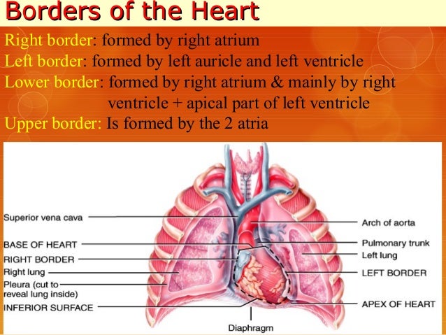
Borders and surfaces of the heart:

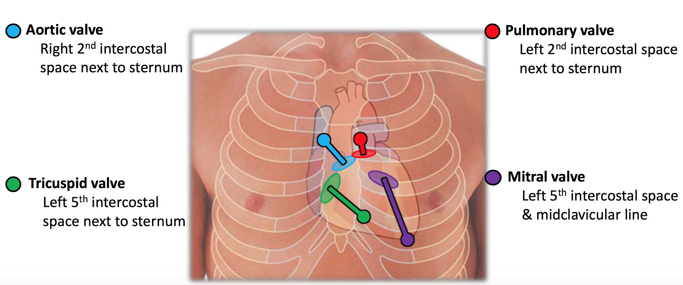
Surface anatomy of the heart:
Blood supply:

Aortic sinus gives off right coronary artery and left coronary artery
Right coronary artery branches:
- Sino arterial nodal
- Right marginal
- AV nodal
- Posterior interventricular
Left coronary artery branches:
- Anterior interventricular
- Circumflex
- Left marginal (from circumflex)
As blood recoils during ventricular diastole, enters coronary arteries to supply heart
NB: Coronary dominance – The coronary artery that supplies SAN, can be right or left or both
Extracardiac anastomosis: Internal thoracic artery branches, bronchial, esophageal, superior and inferior phrenic arteries
Venous:
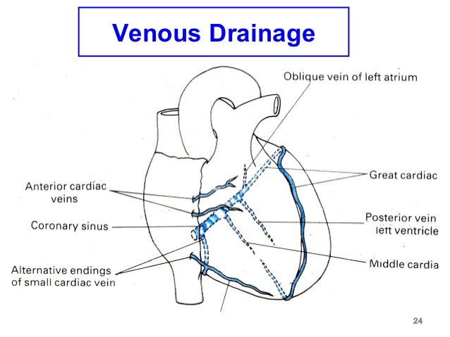
Coronary sinus:
- Runs in posterior coronary groove
- Opens in right atrium between AV orifice and IVC orifice
- Tributaries:
- Great cardiac vein
- Small cardiac vein
- middle cardiac vein
- Left marginal vein
- Left posterior ventricular
- Oblique vein of left atrium
Anterior cardiac veins open directly in right atrium
Lymphatics: Tunica media ⇒ Efferent vessels ⇒ tracheobronchial nodes
Nerves: Sympathetic increases heart rate, parasympathetic decreases
Sympathetic: Right and left cardiac branches of sympathetic ganglia
- Cervical: Superior, middle and inferior ganglia
- Thorax: Ganglia 2,3,4
Parasympathetic:
- Vagus: Right and left upper cervical cardiac branches, right and left lower cervical cardiac branches
- Left recurrent laryngeal – 1 branch
1. Superficial cardiac plexus – Below arch of aorta
- Left superior cervical sympathetic nerve
- Left lower cervical cardiac nerve (parasympathetic)
2. Deep cardiac plexus – infront of tracheal bifurcation
- All the remaining nerves mentioned above

Relations: Same as pericardium
Interior of heart:

(I) Right atrium:
- Crista terminalis (contains SAN)/ Sulcus terminalis – divides atrium into smooth and rough part
- Sinus venarum – smooth – posterior part
- Atrium proper – rough – anterior part
- Pectinate muscles
- SVC, IVC, coronary and AV orifice
(II) Interarterial septum: Fossa ovalis and limbus
Clinicals: Patent foramen ovale
(III) Left atrium:
- Smooth posterior part – absorbed pulmonary veins
- Rough anterior part – Pectinate muscles
(IV) Right ventricle: Divided into 2 by supraventricular crest
- Outflow part – Infundibulum, smooth walls
- Inflow part – Trabeculae carneae which consists of:
- Ridges
- Bridges (eg. moderator band)
- Three Papillary muscles – attached to valves by chorda tendinea – prevents valve prolapse into atria during ventricular systole
(V) Interventricular septum: Superiorly membranous, inferiorly muscular
(VI) Left ventricle:
- Outflow part – Aortic vestibule, smooth walls
- Inflow part – Trabeculae carneae, 2 papillary muscles
Conducting system of the heart:

Triangle of koch: In right atrium, anatomical landmark of AV node
Boundaries: Tendon of Todaro, tricuspid valve and coronary sinus opening

Clinicals:
- Myocardial ischaemia
- Angina pectoris
- Coronary bypass graft – radial artery and long saphenous vein
- Angiogram
- Cardiac referred pain – pain felt in the neck, shoulders, and back
- Heart block 1st, 2nd and 3rd degree
Superior thoracic inlet
Boundaries:
- Anterior – Manubrium
- Posterior – T1 body
- Lateral – 1st rib and costal cartilage
Contents:
- Trachea, esophagus, thoracic duct
- Common carotid artery, subclavian artery and vein, IJV
- Vagus, phrenic, recurrent laryngeal nerves and sympathetic chain
- Apex of lung and pleura
Clinicals: Thoracic inlet syndrome – compression of structures, tumors, enlarged lymph nodes – leads to dysphagia, dyspnea
Inferior thoracic inlet
Boundaries:
- Anterior – 7-10 costal cartilage, xiphisternal joint
- Posterior – T12 body
- Lateral – 11th and 12th ribs
Contents:
- Abdominal aorta
- Azygos vein
- IVC
- Esophagus
- Vagus nerve
- Thoracic duct
Ribcage
(I) Costotransverse and costovertebral joints:

Costotransverse joint – Tubercle of rib articulates with transverse process of corresponding vertebrae
Costovertebral joint – Head of rib articulates with superior costal facet of corresponding vertebrae and inferior costal facet of the vertebra above, as well as the adjacent IVD
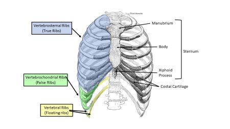
(II) Types of ribs:
(III) Typical rib:
- Anterior – Costal cartilage (hyaline)
- Posterior – Tubercle and head (2 articular facets)
- Superior – Thick and rounded
- Inferior – Sharp, costal groove

(IV) Atypical ribs:


(V) 1st rib relations:
- Superior – clavicle, subclavian vessels
- Inferior – intercostal vessels
- Medial – sympathetic trunk
(VI) Muscles:
1. Intercostal muscles:

- 11 pairs
- Nerve supply – intercostal nerves (T1-T11)
- Intercostal vein, artery and nerve between internal and innermost intercostal muscles
- External – in inspiration elevate ribcage
- Internal – forced expiration
- Innermost – inspiration
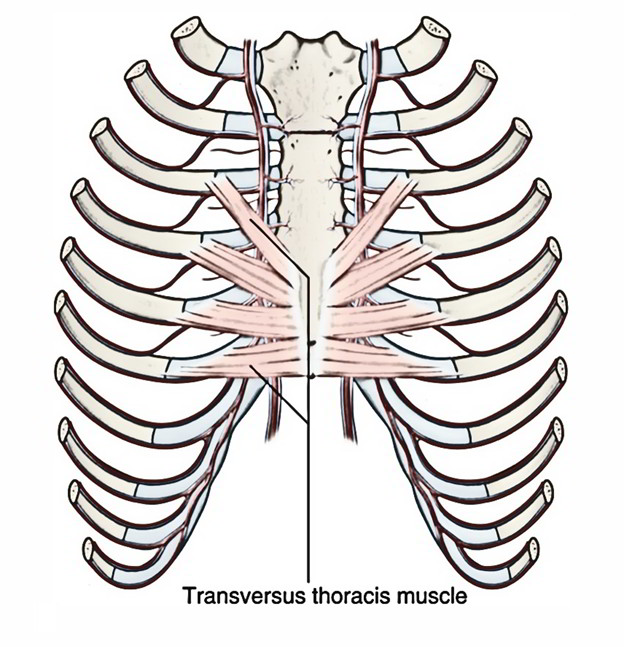
2. Transverse thoracic muscles:
- From posterior inferior sternum to posterior surface of costal cartilage 2-6
- Depress ribs
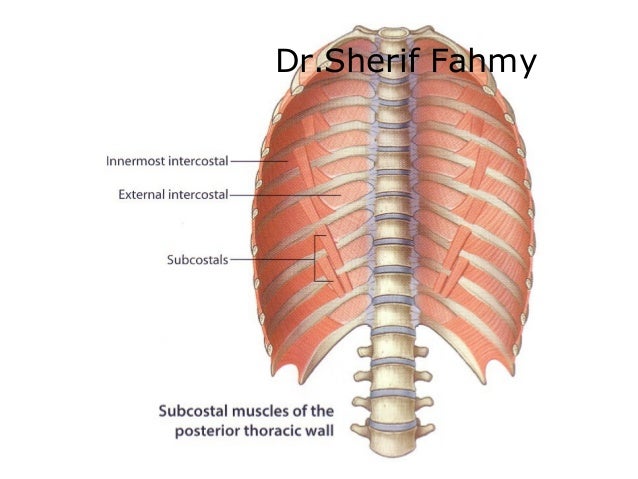
3. Subcostal muscles:
- From posterior lateral rib to a 2nd or 3rd rib below
- Depress ribs
(VII) Muscles of respiration:

(VIII) Thoracic wall/ Ribcage:
1. Blood supply:
- Thoracic aorta – Posterior intercostals, subcostal artery
- Internal thoracic – Anterior intercostals
- Axillary – Superior and lateral thoracic arteries
2. Venous: Azygos system
3. Nerves:
- Supraclavicular nerve – above 2nd rib
- Anterior rami (T1-T11) intercostal nerves
4. Lymphatics: Intercostal, phrenic nodes
Clinicals:
- Age changes – costal cartilage ossify, xiphoid process ossify
- Paralysis of diaphragm, phrenic nerve damaged – paradoxical movement
- Extra ribs – transverse process of cervical or lumbar vertebrae
- Decreased ribs – failure of 12th rib to form
- Rib fracture – at angle or costal cartilage, most common in ribs 3-10 since they are immobile. 1st and 2nd are protected by clavicle, 11th and 12th are mobile.
- Flail chest – anterolateral chest wall movable due to multiple rib fractures. Moves paradoxically (moves outwards during expiration)
- Funnel chest
- Pigeon chest
- Sternal puncture – to get bone marrow from manubrium, pierces skin, fascia and periosteum. May injure aorta, heart, or pericardium
- Median sternotomy – vertical incision along sternum for heart and lung surgeries
Mediastinum
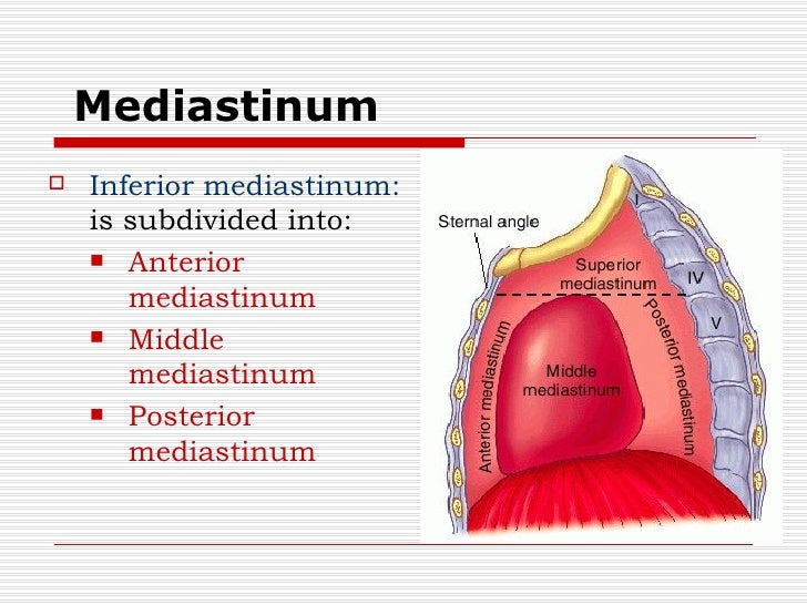
(I) Superior mediastinum:
Relations:
- Superior – Thoracic inlet
- Inferior – Inferior mediastinum
- Anterior – Manubrium
- Posterior – T1-T4
- Lateral – Lungs pleura
Contents:
- Nerves: phrenic, vagus, recurrent laryngeal
- Vessels: SVC, thoracic duct, aorta, internal thoracic artery and vein
- Trachea, esophagus
- Thymus gland
(II) Anterior mediastinum:
Relations:
- Superior – Superior mediastinum
- Inferior – Diaphragm
- Anterior – Sternum
- Posterior – Pericardium
- Lateral – lungs pleura
Contents: Sternopericardial ligament, internal thoracic artery and branches, thymus gland
(III) Middle mediastinum:
Relations:
- Superior – Superior mediastinum
- Inferior – Diaphragm
- Anterior – Pericardium
- Posterior – Pericardium
- Lateral – lungs pleura
Contents: Heart, tracheal bifurcation, phrenic nerve, SVC, pulmonary artery, pulmonary vein, aorta
(IV) Posterior mediastinum:
Relations:
- Superior – Superior mediastinum
- Inferior – Diaphragm
- Anterior – Pericardium
- Posterior – T5-T12
- Lateral – lungs pleura
Contents: Thoracic aorta, thoracic duct, azygos system, esophagus
Vessels and nerves
(I) Internal thoracic artery:
- Originates from 1st part subclavian artery
- Anterior to lung apex
- Enters thorax, posterior to clavicle
- Runs downwards and lateral to sternum
- At 6th intercostal space divides into: superior epigastric (rectus muscle) and musculophrenic (diaphragm)
- Branches: Anterior intercostal arteries, perforators of breast, pericardiophrenic and mediastinal
(II) Aortic arch: (connected to pulmonary trunk by ligament arteriosum)
Location: Sternal angle to lower border T4
Relations:
- Superior – Brachiocepahlic trunk, left common carotid, left subclavian artery
- Inferior – Pulmonary trunk
- Left/anterior – Pleura, phrenic nerve and vagus nerve
- Right posterior – trachea, esophagus
Branches: Brachiocepahlic trunk, left common carotid, left subclavian artery, right and left coronary arteries
(III) Thoracic aorta:
Location: Posterior mediastinum (T4-T12)
Relations:
- Anterior – Pericardium
- Posterior – Vertebral column
- Right – Thoracic duct, azygos vein
- Left – Left lung and pleura
Branches: Posterior intercostals, bronchial, esophageal, pericardial, mediastinal, superior phrenic, subcostal
(IV) Brachiocephalic trunk:
Location: Posterior to manubrium
Relations:
- Anterior – Manubrium
- Posterior – Trachea
- Right – SVC
- Left – Left common carotid
Branches: Right common carotid and right subclavian
(V) SVC:
Extent: 1st-3rd costal cartilage
Location: Anterior and right of superior mediastinum
Relations:
- Anterior – Ascending aorta, right lung
- Posterior – Trachea
- Lateral – Right lung and pleura
Tributaries: Right and left brachiocephalic veins, azygos vein, right and left supreme intercostal veins
(VI) Azygos venous system:
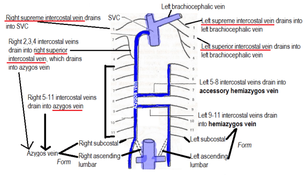
- Azygos vein formed from right subcostal vein and right ascending lumbar vein
- Hemiazygos and accessory hemiazygos drain into azygos vein
- Azygos vein enters thorax via aortic hiatus
- Ascends right of T12 – T4
- Drains into SVC
(VII) Thoracic duct: Main lymphatic trunk
- Continues as cisterna chyli in abdomen
- Enters thorax via aortic hiatus
- In posterior mediastinum, right to thoracic aorta snd posterior to esophagus
- Crossed from right to left at T4
- In superior mediastinum
- Joins junction of left IJV and left subclavian to form left brachiocephalic vein
Territory of drainage: all except superior right quadrant
Clinicals: Laceration – thin wall tears, chyle accumulates in posterior mediastinum
(VIII) Phrenic nerve:
- Origin: Anterior rami of C3,C4,C5
- Begins at lateral border of anterior scalene muscle
- Descends anterior to anterior scalene, deep to prevertebral layer
Right phrenic nerve:
- Passes anterior to 2nd part of subclavian artery
- Enters thorax via superior mediastinum
- Right side of brachiocephalic vein, SVC and pericardium
- Descends anterior to lung root
- Pierce diaphragm near caval opening
Left phrenic nerve:
- Passes anterior to 1st part of subclavian artery
- Enters thorax via superior mediastinum
- Crosses aortic arch and vagus nerve
- Descends anterior to lung root
- Pierce diaphragm
Phrenic nerve distribution:
- Motor and sensory – Diaphragm
- Sensory:
- Parietal pleura
- Parietal pericardium
- IVC
- Suprarenal glands
- Biliary apparatus
Clinicals: Referred pain
(IX) Thoracic sympathetic chain:
- Runs over neck of ribs and transverse process of vertebrae
- Pierce diaphragm to supply abdomen
Trachea, esophagus and thymus gland
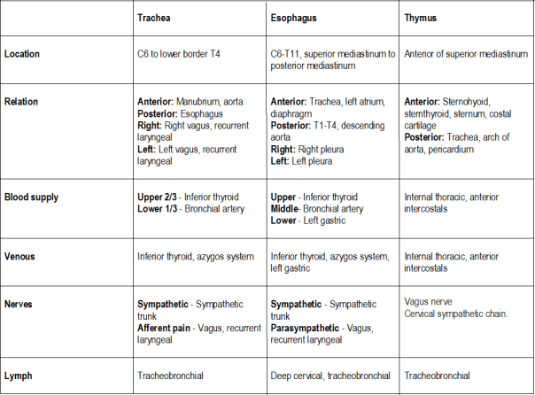
Esophagus:
Constrictors: Cricopharyngeal sphincter, arch of aorta, left main bronchus, diaphragmatic constriction
Clinicals of esophagus: Cancer, compression due to right atrium hypertrophy – dysphagia
Diaphragm
Attachments:
- L1 and L2
- 7-12 rib’s costal cartilage
- Xiphoid process of sternum
- Right (L1-L3) and left (L1-L2) crus – combine to form central tendon
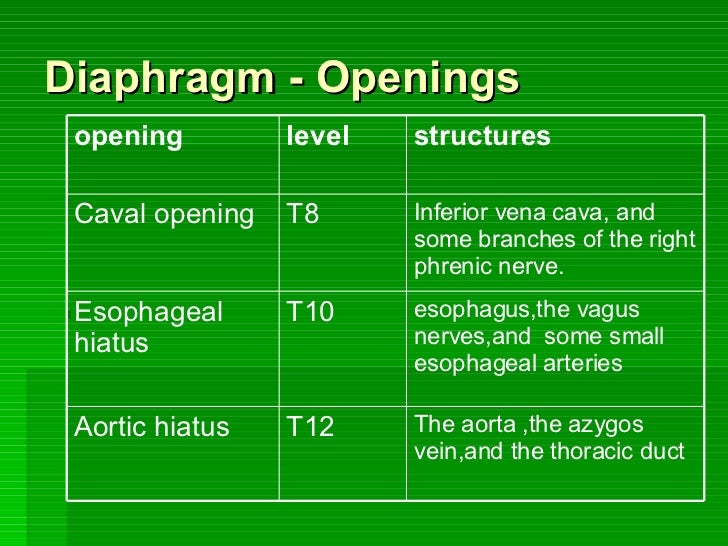
Openings:

Blood supply:
- Internal thoracic – Pericardiophrenic, musculophrenic
- Thoracic aorta – Superior phrenic
- Abdominal aorta – Inferior phrenic
- Lower intercostal arteries
Nerves: Phrenic (motor), intercostal nerves and subcostal nerve (sensory)
Lymphatics: Parasternal, anterior and posterior diaphragmatic
Action:
- Contract, flatten
- Relax, dome shaped
Functions of diaphragm:
- Muscle of inspiration – increase verticle diameter
- Muscle of abdominal straining – helps anterior abdominal muscles to contract, therefore raise intraabdominal pressure for micturition, defecation or parturition
- Weight lifting muscle
- Thoracoabdominal pump – as diaphragm increases intraabdominal pressure and decreases intrathoracic pressure, it compresses blood in IVC and forces it upwards. Thoracic duct also aided.
Relations:
- Superior – Pericardium, lungs
- Inferior – Liver, adrenals, kidney, stomach, spleen
- Posterior – Aorta, azygos vein, esophagus
Clinicals:
- Paralysis (suffocation)
- Hiccups -involuntary contractions of diaphragm, irritation
- Referred pain – shoulder region
- Hiatal hernia – stomach enters thorax via th esophageal hiatus
- Median arcuate ligament syndrome – abdominal pain due to compression of celiac artery
These are summarized notes from various sources, mainly TeachMeAnatomy and Wikipedia
