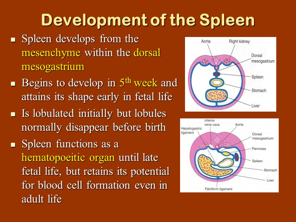
Spleen
Origin: Mesodermal cells in dorsal mesogastrium
- These cells will form stroma and capsule
- Hematopoietic cells infiltrate spleen
- Dorsal mesogastrium forms: gastrosplenic ligament and splenorenal ligament
NB: Hematopoietic function lost with embryo development. Lymphoid precursor cells migrate into developing organ
Congenital anomalies:
- Accessory spleen
- Wandering spleen – lacks one ligament or both
- Polysplenia/ Chaudhrey’s disease – multiple small accessory spleens
Suprarenal glands
- Cortex – Mesodermal cells of intraembryonic coelomic epithelium
- On either side of mesentery of gut, proliferates to form fetal cortex
- Medulla – Sympatho – chromaffin cells from neural crest cells (ectoderm)
- Migrate to enter medial aspect of fetal cortex
- Second layer of cells develop from coelomic mesothelium and surrounds fetal cortex to form permanent cortex
- Fetal cortex regresses and disappears after 3rd year of birth
- Permanent cortex differentiates into 3 zones: zona glomerulosa, zona fasiculata and zona reticularis. Complete histological differentiation attained at puberty.

Congenital anomalies:
- Agenesis
- Ectopic suprarenal gland – below capsule of kidney
- Accessory cortical tissue – found on posterior abdominal wall and pelvis
- Adrenogenital syndrome – hypertrophy of suprarenal cortex and over production of androgens. Results of pseudohermaphroditism in females and premature enlargement of external genitalia in males
