Development of Eyes
(I) Retina and optic nerve
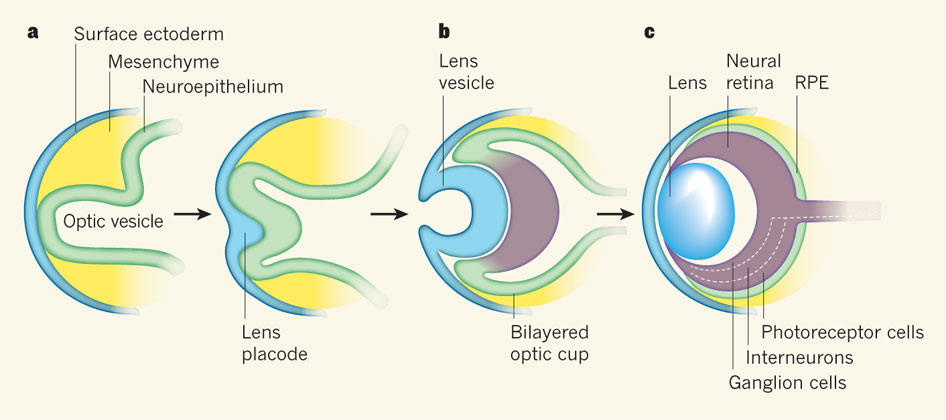
- Optic vesicle of prosencephalon extends laterally
- Contacts surface ectoderm
- Optic vesicle invaginated to form optic cup:
- Outer layer – forms pigmented layer of retina
- Inner layer anterior 1/5 – iris and ciliary body
- Inner layer posterior 4/5 – All layers of retina except pigmented layer
- Mouth of optic cup forms pupil
- Optic stalk invaginated by central artery of retina – gets enclosed in optic stalk – forms optic nerve
(II) Lens
- Surface ectoderm thickens – forms lens placode
- Lens placode invaginated at optic cup mouth – forms lens vesicle
- Lens vesicle separates from surface ectoderm
- Lies in optic cup mouth
- Cells of posterior layer of lens vesicle elongate – forms lens fibers
(III) Choroid and sclera – Forms from mesenchyme around optic vesicle
Development of Ear
Develops from pharyngeal arches (link)
Develops around 4th and 5th week of development, while the middle ear ossicles form around 6 weeks of development.
Muscles of middle ear – tensor tympani (1st arch) and stapedius muscle (2nd arch)
