Origin: Endoderm of foregut
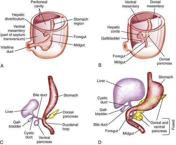
- Liver bud develops from lower end of foregut
- Liver bud divides into two parts:
- Smaller part: Pars cystica ⇒ forms gall bladder
- Large cranial part: Pars hepatica
- Pars hepatica invades septum transversum and divides into right and left branches (right and left hepatic ducts) which branch more to form columns of hepatic cells
- Columns of hepatic cells meet vitelline veins and break them into hepatic sinusoids
- Mesoderm of septum transversum forms fibrous tissue stroma and capsule of liver
- Original stalk of liver bud elongates – forms common bile duct
- Due to rotation of stomach, common bile duct opens in posterior medial part of 2nd duodenum
Ligaments of liver:
- Mesoderm of septum transversum between liver and anterior abdominal wall forms falciform ligament. Umbilical vein lies on inferior free margin of falciform ligament
- Mesoderm of septum transversum between liver and stomach forms lesser omentum
Liver separates from septum transversum except “bare area” of liver
Rest of septum transversum forms part of diaphragm
Congenital anomalies:
- Atresia of common bile duct
- Partial or complete duplication of gall bladder
- Congenital absence of portal vein
- Accessory hepatic duct
