All posts by DentMistry

Development of stomach, duodenum and pancreas

Stomach

Origin: Endodermal

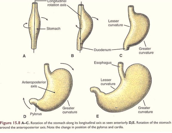
  • Lower part of foregut, swelling appears
  • Anterior border connected to anterior abdominal wall by ventral mesogastrium
  • Posterior border connected to posterior abdominal wall by dorsal mesogastrium
  • Posterior border grows more – forms greater curvature of stomach
  • Anterior border forms lesser curvature
  • Stomach then rotates 90 degrees clockwise (due to growth of liver)
  • So dorsal mesogastrium forms lesser sac
  • Liver develops in ventral mesentery – mesentery becomes lesser omentum
  • Spleen develops in dorsal mesentery – mesentery becomes gastrosplenic ligament

Clinicals: Congenital hypertrophy of pyloric sphincter – narrows pyloric canal. More common in males.

NB: Development of stomach and rotation causes right vagus nerve to become posterior vagus trunk. Left vagus nerve becomes anterior vagus trunk

Image result for development of stomach

Duodenum

Origin: Endodermal

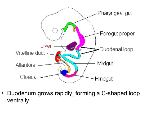
  • Distal foregut and proximal midgut
  • Form a “C” shaped loop projecting ventrally
  • Due to stomachs rotation 90 degrees clockwise
  • Duodenal loop carried dorsally and to the right, becomes adherent to posterior abdominal wall
  • Mesentery of duodenum disappears except in the 1st and 4th part (ligament of trietz)
  • The lumen is temporarily obstructed and canalized again

Congenital anomalies:

  1. Incomplete canalization/ atresia – green vomit, no bowel movement
  2. Incomplete fixation to posterior abdominal wall – sites of internal hernia
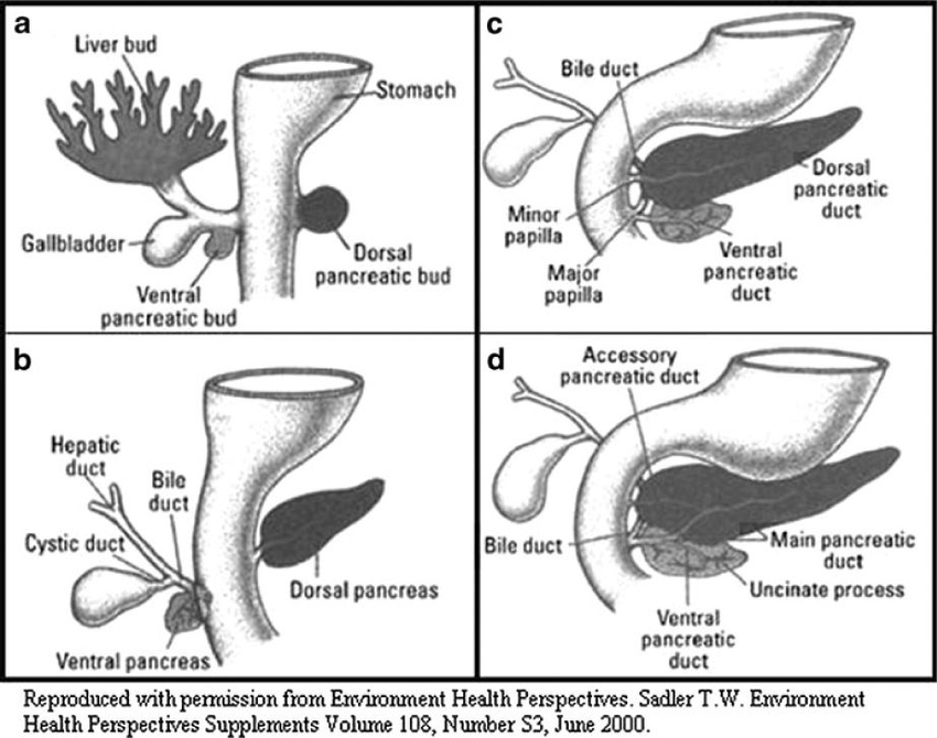
Image result for development of duodenum


Pancreas

Origin: 2 endodermal buds form:

  1. Endoderm of dorsal wall of duodenum – dorsal pancreatic bud
  2. Endoderm of ventral wall of duodenum from hepatic bud stem – ventral pancreatic bud
  • Dorsal pancreatic bud arises slightly above liver bud, extends dorsally and upwards into mesoduodenum
  • Ventral pancreatic bud migrates dorsally to lie below and behind dorsal bud
  • The 2 buds fuse together (ventral bud – head and uncinate process)
  • Ducts of the 2 buds join – main pancreatic duct
  • Islets of Langerhans appear – insulin secretion begins in 5th month

Congenital anomalies:

  1. Annular pancreas – develops as a ring around 2nd part duodenum
  2. Ectopic pancreatic tissue – eg. wall of duodenum, jejunum, ileum or stomach
development of pancreas

Development of eyes and ears

Development of Eyes

Development of eyes

(I) Retina and optic nerve

  • Optic vesicle of prosencephalon extends laterally
  • Contacts surface ectoderm
  • Optic vesicle invaginated to form optic cup:
  1. Outer layer – forms pigmented layer of retina
  2. Inner layer anterior 1/5 – iris and ciliary body
  3. Inner layer posterior 4/5 – All layers of retina except pigmented layer
  • Mouth of optic cup forms pupil
  • Optic stalk invaginated by central artery of retina – gets enclosed in optic stalk – forms optic nerve

(II) Lens

  • Surface ectoderm thickens – forms lens placode
  • Lens placode invaginated at optic cup mouth – forms lens vesicle
  • Lens vesicle separates from surface ectoderm
  • Lies in optic cup mouth
  • Cells of posterior layer of lens vesicle elongate – forms lens fibers

(III) Choroid and sclera – Forms from mesenchyme around optic vesicle


Development of Ear

Develops from pharyngeal arches (link)

Develops around 4th and 5th week of development, while the middle ear ossicles form around 6 weeks of development.

Muscles of middle ear – tensor tympani (1st arch) and stapedius muscle (2nd arch)

Development of CNS (central nervous system)

Origin: ectoderm and neural tube

(I) Neural tubeprevious notes

(II) Neural crest Ectodermal cells on lateral edge of neural tube

Derivatives of neural crest:

  1. All dorsal root ganglia
  2. Autonomic ganglia
  3. Sensory ganglia of cranial nerves V, VII, IX, X (5, 7, 9, 10)
  4. Schwann cells
  5. Medulla of suprarenal gland (chromaffin cells)
  6. Melanoblast cells
  7. Archnoid and pia mater

(III) Spinal cordCaudal part of neural tube

  • Neural tube has a single layer of simple columnar epithelium and central canal
  • The epithelium proliferates – forms 3 layers:
  1. Ependymal layer – produces cerebrospinal fluid (CSF) 
  2. Mantle layer – grey mater
  3. Marginal layer – white mater
development of spinal cord, neural tube

A groove called sulcus limitans divides the mantle layer into:

  1. Alar plate – posterior horn, sensory cells
  2. Basal plate – anterior horn, motor cells

Growth of spinal cord:

  1.  3rd month fetal life – till end of vertebral column
  2. Birth – till L3
  3. Adult – till L1/L2

Congenital anomalies of spinal cord:

  1. Spina bifida oculta – failure of fusion of one vertebrae dorsal part
  2. Meningocele – failure of fusion of 2-3 vertebra. meninges bulge out
  3. Meningo-myelocele – spinal cord bulges out
  4. Myelocele – Neural tube failed to close, neural plate exposed
Image result for meningomyelocele vs meningocele
Image result for myelocele
congenital anomalies of spinal cord

(IV) BrainCranial end of neural tube

  • Neural tube expands to form brain swelling
  • Lumen forms ventricles
  • 2 constrictions divide brain swelling into 3 parts

(V) Cerebellum Alar lamina of metencephalon

  • Alar lamina bend medially, forming medial and lateral bulges
  • The medial bulges meet each other over the roof plate of 4th ventricle, forming vermis 
  • Lateral bulge forms cerebeller hemispheres
  • Cerebeller cortex formed by – neuroblast cells migrating from mantle layer to marginal layer
  • Dentate nucleus – neuroblasts deeply situated in mantle layer
  • Cerebeller peduncles – axons of neurons of cerebeller nuclei grow out to reach brainstem
Image result for development of cerebellum

Development of vertebral column

  • Notochord forms
  • Sclerotome cells (paraxial mesoderm) surround notochord
  • Form mesenchymal vertebral column

Image result for caudal half of one sclerotome

  • Caudal half of one sclerotome and cranial half of the next, forms one vertebral mesenchyme, which goes through endochondral ossification  to form bone
  • Notochord regresses in regions of vertebral body
  • Notochord enlarges between the bodies to form nucleus pulposus

Image result for caudal half of one sclerotome notochord


Each vertebral body gives 3 processes:

  • 2 dorsal process – unite to form spinous process
  • 2 lateral process – in cervical region, unites with coastal process to form foramen transversarium. In thoracic region forms transverse process
  • 2 coastal process – in thoracic region elongates to form ribs

Image result for cervical and thoracic vertebrae


Development of mammary glands

  • Linear thickening of surface ectoderm (milk line) appears on ventral body
  • Extends from axilla to medial inguinal region
  • Line disappears except in pectoral region
development of mammary glands, milk line

  • 15 to 20 solid ectodermal buds arise in remaining milk line
  • Buds form solid cords
  • Which grow in underlying mesenchyme
  • Cord grows and branches in fetal life
  • Canalized before birth to form: Lactiferous sinus, Lactiferous duct, Secretory alveoli
  • Open in depressed area, which elevates after birth – to form the nipple
Image result for lactiferous sinus

At puberty:

  • Males: have ducts without alveoli
  • Females: Ovarian hormone increases branching of lactiferous duct, and increases fat between lobules. Therefore the mammary gland enlarges.

Congenital anomalies:

  1. Amastia – absence of one or two mammary glands
  2. Athelia – absence of one or two nipples
  3. Macromastia – large breasts
  4. Micromastia – small breasts
  5. Polythelia – accessory nipples
  6. Polymastia – accessory breast
  7. Gynaecomastia – male developing female breasts
  8. Klinefelter syndrome – XXY in males, therefore have enlarged breasts, wide hips and penis does not grow

Development of skin

The development of skin consists formation of:

  • Epidermis
  • Dermis
  • Subcutaneous tissue
  • Hair
  • Sweat glands
  • Sebaceous glands

(I) Epidermis:

  • Ectoderm
  • Neural crest cells – Form melanoblasts (melanin secreting cells)

(II) Dermis – paraxial mesoderm (dorsal portion) – dermatome (contributes skin, fat and connective tissue)

(III) Subcutaneous tissue – lateral plate mesoderm – somatic layer (most of the skin derived)

(IV) Hairs:

  • Solid ectodermal buds sink into underlying mesenchyme
  • Forms hair bud, which is invaginated by mesoderm
  • Forms hair papilla
  • Muscle fibers attach to hair follicles – errector pili muscles
  • Hair bulb central cells elongate and keratinize – hair shaft
  • Fetus first hair – lanugo hair
Image result for Development of hair

(V) Sweat glands:

  • Solid ectodermal buds sink into underlying mesenchyme
  • Terminal end coils and it canalizes
Image result for Development of sweat glands

(VI) Sebaceous glands:

  • Side branches of hair follicle buds
  • Central cells degenerate forming fatty secretions

Congenital anomalies:

  1. Ichthyosis – dry scaly skin
  2. Hypertrichosis – excessive hair
  3. Atrichia – congenital absence of hair
  4. Dermoid – cystic swelling along lines of fusion

Development of skeletal muscles and limbs

Skeletal muscles

Origin: Paraxial mesoderm – myotomes

Myotomes ⇒ myoblast cells ⇒ fuse ⇒ myotubes ⇒ myofilaments develop in myotubes ⇒ myotubes become myocytes (muscle cells)

somites, development of head and neck, development of skeletal muscles and limbs
  • 1st occipital myotome – extra occular muscles of the eye (NB: iris and ciliary muscle – neural ectoderm)
  • Remaining 3 occipital myotomes – muscles of tongue


somite development

The 8 cervical, 12 thoracic, 5 lumbar, 5 sacral, 8-10 coccygeal myotomes divide into:

  • Dorsal part – Epimere – forms extensor muscles of back and neck
  • Ventral part – Hypomere:
  1. Split longitudinally eg. infrahyoid muscles
  2. Split to form layers eg. intercostal muscles
  3. Ventral tips unite to form eg. rectus abdominis muscle
development of skeletal muscles and limbs

NB:

  • Myotomes migrate to limb buds to form flexor and extensor muscles
  • Pharyngeal arches give muscles in head and neck

Limbs

  • Upper limb bud forms opposite C5-T1
  • Lower limb bud forms opposite L4-S3

Each limb bud is formed of:

  • Lateral plate mesoderm – somatic layer – bones, tendons and connective tissue
  • Migrating myotomes – muscles
  • Neural crest cells – melanocytes and schwann cells
  • Which is all covered by ectoderm – skin

Development:

  • Programmed apoptosis between digital rays form fingers and toes
  • Central mesoderm – cartilagenous skeleton – ossify into bone
  • 3 segments formed in each limb bud
  • Rotation of 90 degrees around long axis:
  1. Upper limb adducted, rotated laterally, thumb lateral
  2. Lower limb adducted, rotated medially, big toe is medial

Congenital anomalies of limbs:

  1. Amelia – failure to develop limbs
  2. Phocomelia – absence of proximal limbs, direct foot or hand
  3. Polydactyl – Extra fingers or toes
  4. Syndactyl – fusion of 2 fingers or toes
  5. Claw feet – lobster deformity
  6. Congenital digit amputation
  7. Constriction band
  8. Club foot
  9. Ectodactyl – missing middle finger

Development of face and palate

Face

5 processes formed by proliferation of neural crest cells (ectoderm)

These cells migrate from mesencephalon and rhombencephalon into arches

  • 1 frontonasal process – cranial to stomodeum
  • 2 maxillary processes – lateral, from 1st pharyngeal arch
  • 2 mandibular processes – caudal, from 1st pharyngeal arch

1 frontonasal process:

  • 2 nasal placodes develop
  • Each invaginated to form:
  1. Nasal pit – forms nasal cavities – open in pharynx posteriorly
  2. Lateral nasal fold – ala of nose
  3. Medial nasal fold – fuse together to form:
  • Surface: middle part of nose, filtrum of upper lip
  • Deep: anterior upper jaw with Incisor teeth, primary palate with incisive fossa

NB: Also forms nasal septum

2 maxillary processes:

  • Grow medially
  • Fuse with medial nasal fold – form upper lip
  • Fuse with lateral nasal fold – form nasolacrimal duct
  • Fuse with mandibular process – forms cheeks
  • Forms palatine shelves

2 mandibular processes:

  • Fuse with each other medially – form lower lip and chin
  • fuse with maxillary process – forms cheeks

NB: Degree of fusion of max. and mand. processes determine width of mouth

Image result for development of face


Palate

Origin: Neural crest cells (ectoderm)

  • Formed by primary and secondary palate
  • Primary palate formed from frontonasal process (fusion of medial nasal folds)
  • Secondary palate formed from 2 palatine shelves, which arise from maxillary process
  • 2 palatine shelves fuse with each other medially, and primary palate anteriorly
  • Nasal septum descends downwards and fuses with palate in midline
  • Anterior part of palate ossifies and posterior part remains fleshy to form hard and soft palate

Anomalies of face and palate:

  1. Inclusion dermoid – cystic swellings around lines of fusion of face
  2. Macrostomia – large mouth, incomplete fusion between max. and mand. processes
  3. Microstomia – small mouth, excessive fusion between max. and mand. processes
  4. Oblique facial cleft – failure of fusion between maxillary and frontonasal process
  5. Lateral cleft upper lip – unilateral or bilateral
  6. Cleft palate – unilateral, bilateral, only soft palate or cleft uvula

Development of respiratory system

Development of larynx, trachea, lungs and esophagus

Respiratory system

Origin: Endodermal floor of pharynx behind hypobranchial eminence

1. Epithelium

  • Laryngo-tracheal groove appears in the endodermal floor of pharynx behind hypobranchial eminence
  • Edges of groove unite dividing upper foregut into esophagus and laryngo-tracheal tube
  • Laryngo- tracheal tube grows caudally:
  1. Upper end – larynx
  2. Then – trachea
  3. Lower part – 2 lung buds
  • Right lung bud divides into 3 branches (main bronchii)
  • Left  lung bud divides into 2 bronchii
  • Each bronchus – subdivisions – bronchial tree
  • Terminal/respiratory bronchioles end in alveoli, expand only after birth

2. Cartilage, muscles and connective tissue: 4th and 6th arch mesoderm

  • 4th pharyngeal arch  – thyroid cartilage, cricothyroid muscle
  • 6th pharyngeal arch – cricothyroid, arytenoid, cuneiform and corniculate cartilages, and all other laryngeal muscles

NB: Splanchnic and somatic pleura forms lungs viceral and parietal pleura

Image result for laryngotracheal groove

Maturation of lungs:

Image result for maturation of lungs embryology
Image result for maturation of lungs
embryology

Anomalies of lungs:

  • Agenesis of one or both
  • Accessory lung lobe
  • Hyaline membrane disease – alveoli cannot ventilate adequately due to absence of surfactant

Esophagus

Origin: Endoderm of foregut

  • From respiratory diverticulum to stomach swelling
  • Surrounding mesenchyme forms musculature of esophagus

Anomalies of trachea and esophagus:

  1. Tracheal-esophageal fistula – incomplete fusion of laryngo-tracheal groove
  2. Esophageal atresia – narrowing of esophagus
  3. Failure of elongation – pulls stomach up thorax – Hiatus hernia

Development of tonsils, tongue and thyroid

Development of palatine tonsils, tongue and thyroid gland

Palatine tonsils

Origin: 2nd pharyngeal arch endoderm

Endoderm proliferates into buds ⇒ Canalizes ⇒ Forms crypts ⇒ Invaded by lymphocytes

Development of palatine tonsils

Tongue

  1. Muscles of tongue – 3 occipital myotomes of paraxial mesoderm
  • (1st occipital myotome forms extraoccular muscles of eye)
  • The 3 remaining myotomes drag the hypoglossal nerve with them

2. Mucous membrane:

  • Anterior 2/3 – 1st pharyngeal arch
  • From two lingual swellings and tuberculum impar
  • Posterior 1/3 – endoderm of 3rd pharyngeal arch
  • Endoderm of 2,3 and 4 arch fuse to form Hypobronchial eminence
  • A groove divides the hypobronchial eminence into:
  1. Upper part – Posterior 1/3 tongue mucous membrane
  2. Lower part – Forms epiglottis

Anterior and Posterior fuse at sulcus terminalis ( v shape)

Development of tongue

Congenital anomalies:

  • Aglossia – absence of tongue
  • Macroglossia, microglossia
  • Bifid tongue
  • Glossoschisis – cleft tongue
  • Tongue tie (ankyloglossia) – frenulum is till tip of tongue, can’t protrude tongue

Thyroid gland

  1. Endodermal thickening around foramen cecum
  2. Forms thyroid primodium between tuberculum impar and hypobronchial eminance
  3. Thyroid primodium invaginated – bilobed diverticulum
  4. Descends infront of hyoid bone and laryngeal cartilage
  5. Connected to dorsum of tongue by thyroglossal duct
  6. Finally reaches infront of thyroid cartilage and upper trachea
  7. Thyroglossal duct obliterated
Image result for development of thyroid gland

Congenital anomalies:

  • Thyroid agenesis – absence, leads to stunted physical and mental growth
  • Lingual thyroid – Fails to descend
  • Thyroglossal cyst – thyroglossal duct unobliterated
  • Thyroglossal fistula – cyst may rupture
  • Ectopic positions – Sublingual, retrohyoid, retrosternal etc.