Arteries
1. Blood supply of face:

- External carotid artery branches ^^
- Ophthalmic artery branches: supratrochlear, supraorbital
2. Ophthalmic artery:
- 1st branch of internal carotid artery
- Through optic canal
- Runs in medial wall of orbit
- Branches:
- Lacrimal
- Central retinal
- Supratrochlear
- Supraorbital
3. Maxillary artery:
- Branch of external carotid artery, arises behind neck of mandible
1st part (mandibular):
- Passes between mandible ramus and sphenomandibular ligament
- Branches:
- Deep auricular
- Anterior tympanic
- Inferior alveolar
- Middle meningeal
- Accessory meningeal
2nd part (pterygoid):
- Passes between 2 heads of lateral pterygoid muscle and enters pterygoid fossa
- Branches:
- Masseteric
- Deep temporal
- Pterygoid
- Buccal
3rd part (pterygomaxillary):
- Lies in pterygopalatine fossa
- Branches:
- Sphenopalatine artery
- Greater and lesser palatine arteries
- Posterior superior alveolar artery
- Pharyngeal artery
- Infraorbital artery
4. Facial artery:
- Emerges in carotid triangle from external carotid artery (ECA)
- Deep to mandible ramus
- Superficial to masseter and buccinator
- Ascends lateral nose
- Becomes angular artery
- Branches:
- Superior labial
- Inferior labial
- Lateral nasal
- Angular
5. Subclavian artery:
- Right one arises from brachiocephalic artery, left one arises from arch of aorta
- It is divided into 3 parts as it passes posterior to anterior scalene muscle
- Branches:
- 1st part – Vertebral, internal thoracic, thyrocervical
- 2nd part – Superior intercostal, deep cervical
- 3rd part – Dorsal scapular
- Continues as axillary artery at border of 1st rib
6. Common carotid artery:
- Right from brachiocephalic trunk, left from arch of aorta
- Bifurcates into ECA and ICA at superior border of thyroid cartilage
7. External carotid artery:
- Formed from common carotid artery
- At upper border of thyroid cartilage
- Outside carotid sheath
- Posterior to ramus of mandible
- Terminates as superficial temporal and maxillary artery
8. Vertebral artery:
- From subclavian artery 1st part
- Through vertebral triangle
- Ascend in transverse foramina C6-C1
- Enter cranial cavity via foramen magnum
- Joins other side’s vertebral artery to form basilar artery at base of pons
Veins
1. Venous drainage of face

2. Facial vein:
- Tributaries: Supraorbital and supratrochlear drain into angular vein
- Becomes facial vein
- Superficial to masseter, buccinator and mandible
- Joins anterior division of retromandibular vein
- To form common facial vein
- Drains into IJV
3. External jugular vein (EJV):
- Formed from retromandibular vein, posterior division and posterior auricular vein
- Forms at angle of mandible
- Pierce deep fascia
- Drain into subclavian vein
4. Internal jugular vein (IJV):
- Formed from sigmoid sinus and inferior petrosal sinus
- Through jugular foramen
- Runs in carotid sheath
- Unites with subclavian vein to form brachiocephalic vein
- Tributaries: Common facial, lingual, pharyngeal, superior and middle thyroid veins
5. Subclavian vein:
- Continuation of axillary vein from border of 1st rib
- Anterior to scalenus anterior muscle
- Joins IJV and EJV to form brachiocephalic vein
Nerves
1. Nerve supply to face:
- Motor – facial nerve branches
- Sensory – trigeminal nerve and nerves C2, C3, C4
2. Inferior alveolar nerve:
- Branch of V3
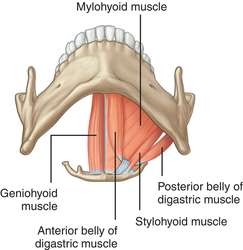
- Gives off a branch – mylohyoid nerve (mylohyoid and anterior diagastric muscle)
- Between mandible ramus and medial pterygoid muscle
- Enters mandible foramen, through mandible canal
- Through inferior dental plexus
- Gives off a mental nerve (at mandibular 2nd premolar) which exits via mental foramen (sensory to chin and lower lip)
- Continues as mandibular incisive nerve to innervate mandibular canines and incisors
Clinicals:
- Inferior alveolar nerve block – anesthesia near mandibular foramen
- Injury – 3rd molar removal, dental implants, root canal
3. Lingual nerve:
- Branch of V3
- Chorda tympani nerve (of facial nerve) joins lingual nerve
- Between mandible ramus and medial pterygoid muscle
- Inferior to 3rd molar
- Runs between hyoglossus muscle and deep part of submandibular gland
- Crosses lateral to medial over Wharton’s duct
- Runs along tip of tongue becoming sublingual nerve, lying beneath mucous membrane
Clinical: 3rd molar surgery – injury to nerve
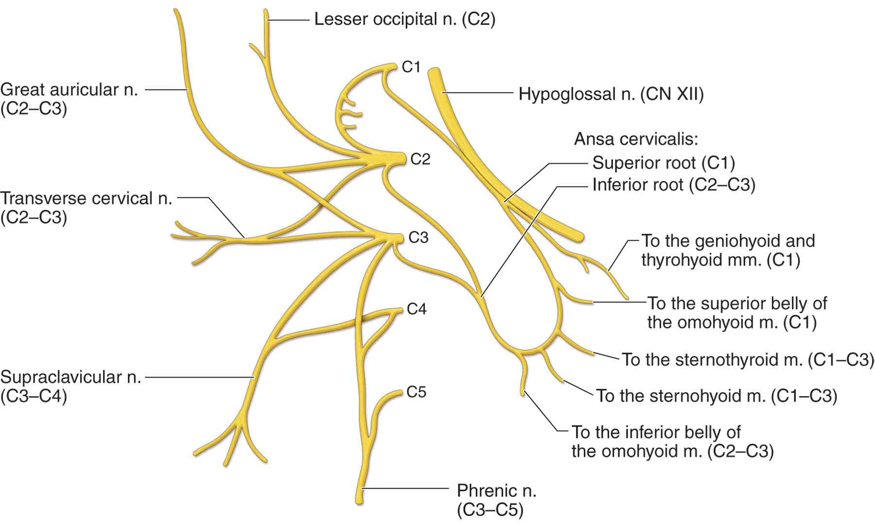
4. Cervical plexus:
Anterior rami C1-C4 – in carotid triangle
5. Gustatory pathway:

Waldeyer’s ring

Palatine tonsils:
- Location: Between palatoglossus and palatopharyngeus folds
- Relations:
- Anterior – palatoglossus fold
- Posterior – palatopharyngeus fold
- Superior – soft palate
- Inferior – tongue
- Lateral – superior constrictor
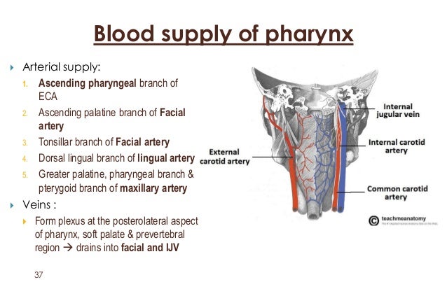
- Blood supply:
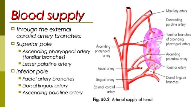
- Nerve: Glossopharyngeus nerve, lesser palatine nerve (V2)
Clinicals: Tonsillitis, tonsilectomy
Temporomandibular joint (TMJ)
Lateral pterygoid muscle attatches to TMJ capsule – slide forward movement
Classification: Synovial modified hinge
Lined by: Fibrocartilage
Articular surfaces: Condyle, mandibular fossa and articular tubercle of squamous temporal

Stability factors:
Static:
- Mandibular fossa and posterior glenoid tubercle
- Articular disc – attaches to internal surface of joint capsule, dividing it into superior and inferior cavity
- Condyle head more convex antero-posteriorly than medial to lateral
- Lateral pole more anterior than medial
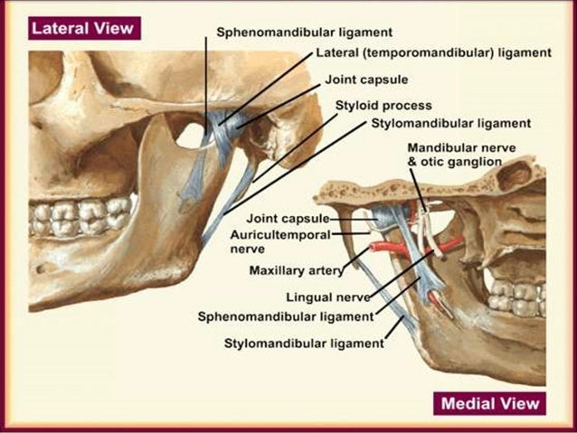
- Ligaments:
- Lateral ligament – from articulating eminence to posterior condyle, prevents extreme retrusion
- Lateral and medial collateral ligament
- Sphenomandibular ligament – from sphenoid spine to lingula, prevents extreme protrusion
- Stylomandibular ligament – from styloid process to angle of mandible
Dynamic: Muscles of mastication
Blood supply: Superficial temporal and masseteric arteries
Nerve supply: Auriculotemporal and masseteric
Movements: Rotation, Protraction
Relations:
- Anterior – lateral pterygoid muscle
- Posterior – parotid gland
- Lateral – parotid gland
- Medial – spine of sphenoid
- Superior – middle cranial fossa
- Inferior – maxillary artery
Glands
(I) Lacrimal gland:
Blood supply: Lacrimal artery from opthalmic artery
Nerve supply:

(II) Parotid gland:
Relations:
- Superior – zygomatic arch
- Inferior – mandible angle
- Anterior – masseter muscle
- Posterior – sternocleidomastoid muscle (SCM)
- Roof – skin and fascia
- Floor – masseter, SCM, mandible ramus
Stenson’s duct course: Anterior to masseter, pierce buccinator, open in vestibule next to 2nd maxillary molar
Pierced by: Superficial temporal artery, retromandibular vein, facial nerve
Blood supply: Superficial temporal artery
Venous: Retromandibular vein
Nerve supply:

- Parasympathetic: Lesser petrosal nerve
- Sympathetic: Superior cervical ganglion
Lymphatic drainage: Posterior and preauricular lymph nodes
Type of secretion: Serous
Clinicals:
- Parotid gland tumor
- Parotiditis – inflammation
- Mumps
(III) Submandibular gland:
Location: Submandibular triangle
Relations:
- Superior – mylohyoid line
- Inferior – diagastric tendon
- Anterior – mental foramen
- Posterior – mandible angle
- Medial – root of tongue
- Lateral- masseter and mandible
Wharton’s duct course: Through 3 muscles (mylohyoid, hyoglossus, genioglossus) ⇒ crossed by lingual nerve ⇒ opens near frenulum
Nerve supply:
- Parasympathetic: Vasodilation
- Sympathetic: Vasoconstrict, therefore enzyme rich mucous
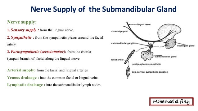
Lymphatic drainage: Submandibular lymph nodes, which drain to jugulodiagastric lymph nodes
Type of secretion: Serous and mucous (seen as demilunes in histology)
Clinicals:
- Submandibular excision – damage lingual and facial nerve
- Calcified stones – due to ascending duct, serous and mucous secretions, and it’s a long duct
(IV) Sublingual gland:
Location: Sublingual fossa above mylohyoid line
Relations:
- Superior – mucous membrane of mouth
- Inferior – mylohyoid muscle
- Posterior – submandibular gland
- Medial – genioglossus muscle
- Lateral- sublingual fossa
Blood supply, venous drainage, nerve supply and lymph nodes – same as submandibular gland
Type of secretion: Mucous – sublingual papilla
Clinicals:
- Ranula – mucous cysts in floor of mouth
(V) Thyroid gland:
Location: Anterior neck, below laryngeal prominence
Extent: C5-T1
Relations:
- Anterior – sternohyoid, sternothyroid
- Posterior – trachea
- Superior – cricothyroid cartilage
- Inferior – 5 tracheal rings
- Medial – esophagus
- Lateral – carotid sheath
Blood supply: Superior, middle, inferior thyroid artery and vein
Nerves:
- Sympathetic: Cervical sympathetic ganglions (superior, middle, inferior)
- Parasympathetic: Vagus nerve
Lymphatics: Pretracheal, paratracheal and prelaryngeal lymph nodes

Clinicals:
- Goiter – enlarged thyroid gland
- Thyroidectomy – surgical removal
- Tracheotomy – forming an opening into trachea due to sudden obstruction of vital airways
- Laryngoscopy
Muscles
(I) Extraocular muscles:

Blood supply: Ophthalmic artery
Nerve supply: Oculomotor, Trochlear (superior oblique), Abducens (lateral rectus)
(II) Muscles of mastication:
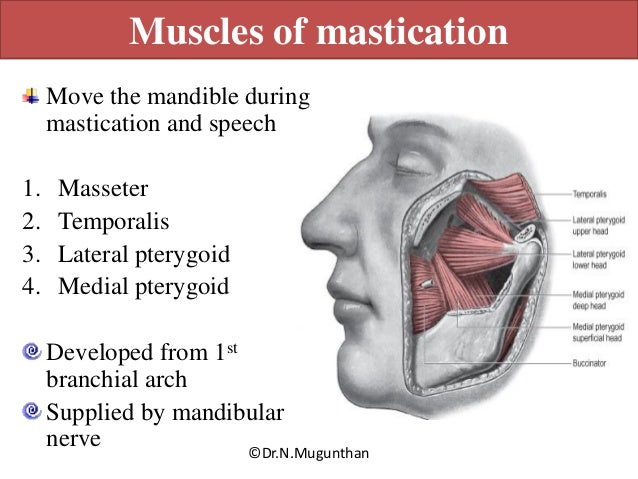

(III) Suprahyoid muscles:

(IV) Infrahyoid muscles:


(V) Sternocleidomastoid (SCM):
- Origin: 2 heads – manubrium, medial 1/3 clavicle
- Insertion: mastoid process
- Innervation: CN 11
- Action: Turn head opposite side, raise thorax
- Relations:
- Anterior – platysma muscle
- Posterior – carotid sheath
- Medial – ansa cervicalis
- Lateral – subclavian artery
(VI) Scalenus anterior:
- Origin: Transverse process C3-C6
- Insertion: 1st rib, scalene tubercle
- Innervation: Anterior rami C4-C6
- Relations:
- Anterior – SCM, subclavian vein
- Posterior – 2nd part subclavian artery, brachial plexus
- Medial – 1st part subclavian artery
- Lateral – 3rd part subclavian artery, brachial roots
Clinicals: Scalenus anterior syndrome – hypertonic muscle, compresses structures
Spaces
(I) Orbit:
Boundaries:

Foramens/fissures and their contents:

Orbit contents: Extraocular muscles and ciliary ganglion
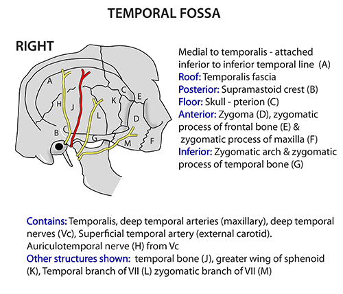
(II) Temporal fossa:
(III) Infratemporal fossa:

Contents: Lateral and medial pterygoid muscles, maxillary artery, mandibular nerve, otic ganglion
(IV) Pterygopalatine fossa:


Clinicals: Ligate sphenopalatine artery to stop nose bleeding
(V) Neck triangles:
(A) Anterior neck triangles
Boundaries:

NB: Submandibular, Carotid and Muscular triangle are paired. Submental triangle is the only unpaired triangle.
Contents:
Submental triangle:
- Submental lymphnodes
Submandibular (diagastric) triangle:
- Hypoglossal nerve
- Nerve to the mylohyoid
- Marginal mandibular branch of the facial nerve (MMB)
- Facial and lingual arteries and veins
- Submandibular gland
- Lower pole of the parotid gland
- Submandibular lymph nodes
Carotid triangle:
- Common carotid artery (and its bifurcation into ECA & ICA)
- Arteries: Superior thyroid, lingual, facial, occipital, and ascending pharyngeal arteries
- Veins: Superior thyroid, lingual, facial, ascending pharyngeal, and occipital veins – Drain into IJV
- Nerves: Hypoglossal nerve, the external and internal branches of the superior laryngeal nerve arising from the vagus nerve
Muscular triangle:
- Muscles: Sternohyoid, sternothyroid, omohyoid, and thyrohyoid muscles
- Superior thyroid artery
- Anterior jugular and inferior thyroid veins
- Ansa cervicalis
- Anterior cervical, infrahyoid, prelaryngeal, thyroid, pretracheal, paratracheal lymph nodes
- Medial part: Esophagus, trachea, thyroid gland, and the lower part of the larynx.
(B) Posterior neck triangles
Boundaries:

- Roof: Investing fascia
- Floor: Anterior, middle and posterior scalene muscles
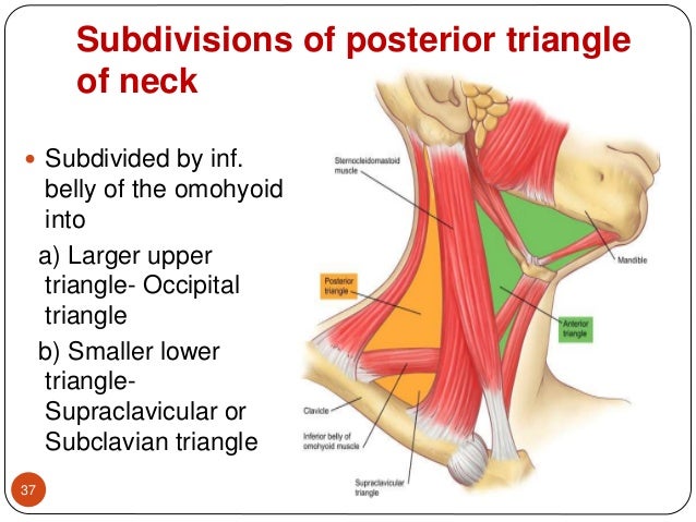
Contents:
- Occipital: Brachial roots, cervical plexus, occipital artery
- Subclavian: Subclavian artery and vein, EJV, brachial plexus trunks
(VI) Interscalene triangle:
- Boundaries: Anterior scalene, middle scalene, 1st rib
- Contents: Subclavian artery, brachial plexus roots
(VII) Suboccipital triangle:
Boundaries:

Contents: Vertebral artery, suboccipital venous plexus
Clinicals: Angiography of circle of Willi’s
Nasal cavity:

Extent: Vestibule to nasopharynx
3 parts: Vestibule, olfactory region, respiratory region
Functions:
- Humidify air – rich vascular supply
- Conchae slow down air
- Prevent pathogens
- Smell
- Drain paranasal sinus
Boundaries:
- Superior – ethmoid and sphenoid bone
- Inferior – palatine bone
- Medial – septa
- Lateral – conchae
Below superior, middle and inferior conchae are meati (openings)

Openings in the meati:
- Above superior conchae/ sphenoethmoidal recess – Sphenoidal sinus
- Superior meatus – Posterior ethmoidal sinus
- Middle meatus – Frontal, maxillary and anterior ethmoidal sinus
- Inferior meatus – Auditory tube, nasolacrimal duct
Other openings:
- Cribriform plate – olfactory nerves
- Sphenopalatine foramen (connects pterygopalatine fossa) – sphenopalatine artery, nasopalatine nerve
- Incisive canal (connects oral cavity) – nasopalatine nerve to oral cavity
Blood supply:
- Internal carotid artery: Anterior and posterior ethmoidal via cribriform plate
- External carotid artery:
- Maxillary artery – Sphenopalatine, greater palatine
- Facial artery – Superior labial, lateral nasal
Venous: Ophthalmic vein, angular vein, sphenopalatine vein
Nerves:
- Smell – olfactory
- General sensory – nasopalatine, nasociliary (V2)
Clinicals: Spread of respiratory infection to ear
Paranasal sinuses:

- Air filled extensions of nasal cavity
- Reduce weight of skull
- Humidify air
- Are paired
- Are mucous lined

Clinicals:
- Sinusitis
- Inflammation of maxillary sinus – tooth ache
- Rhinitis – inflamed nasal mucosa
- Epistaxis – nose bleed (trauma or hypertension)
Tongue:
4 types of papillae:

- Filiform – all over the tongue, only type that contain no taste buds
- Fungiform – mushroom shaped, concentrated on tip of tongue
- Foliate – found on postero-lateral surface of tongue
- Circumvallate – 12 to 14, anterior to sulcus terminalis, Von Ebner’s glands open in it
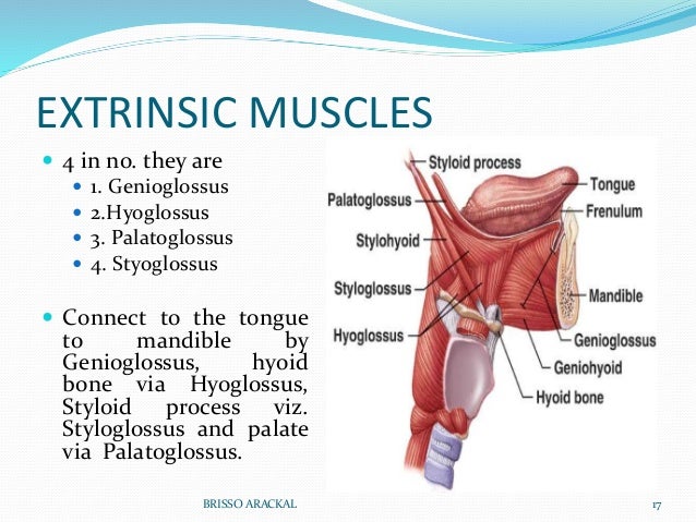
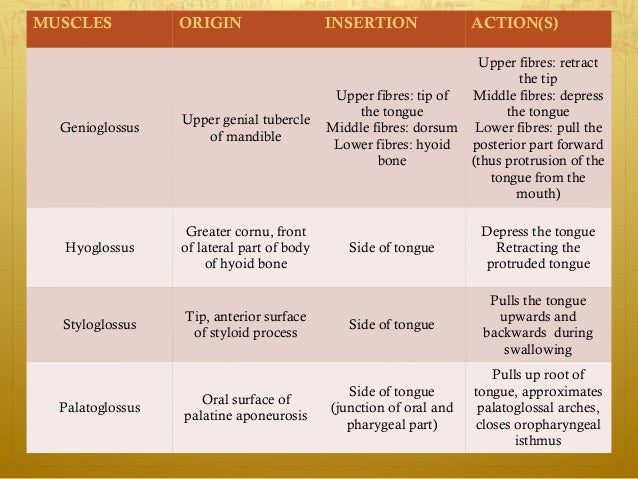
Extrinsic muscles:
Intrinsic muscles:


Blood supply: Lingual artery and vein
Nerve supply:
- Taste: Anterior 2/3 chorda tympani nerve, Posterior 1/3 glossopharyneal nerve
- General sensation: Anterior 2/3 Lingual nerve (V3), Posterior 1/3 glossopharyneal nerve
- Motor: Hypoglossus nerve, except palatoglossus muscle which is supplied by vagus nerve
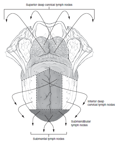
Lymphatic drainage: Superior deep cervical, inferior deep cervical, submandibular, submental lymph nodes
Clinicals:
- Tongue tie
- Cancer
- Halitosis – bad breath
- Oral thrush
Hard palate and soft palate
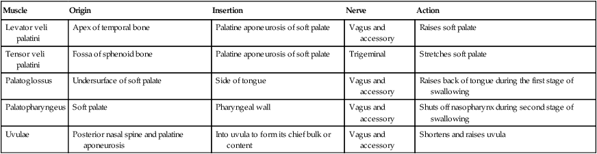
Soft palate muscles:
Blood supply:
Hard palate: Greater palatine artery (from descending palatine)
Soft palate:
- Greater/ lesser palatine (from maxillary artery)
- Ascending palatine (from facial artery)
- Ascending pharyngeal (from ECA)
Veins: Drain into pterygoid venous plexus
Nerves:
Hard palate:
- Greater palatine – mucosa of posterior hard palate (from pterygopalatine ganglion, descends through greater palatine foramen with greater palatine artery)
- Nasopalatine – anterior mucosa (through incisive foramen)
Soft palate:
- Pharyngeal plexus via vagus nerve
- Except tensor veli palatini – medial pterygoid nerve (V3)
- Sensory – Lesser palatine nerve
Lymphatic drainage:
Hard palate: Submandibular, superior deep cervical nodes
Soft palate: Retropharyngeal, superior deep cervical nodes
Clinicals:
- Cleft palate
- Palatal abscesses
- Cleft uvula
- Pimples on hard palate
Pharynx
Extent: Base of skull to C6
Layers:
- Buccopharyngeal fascia
- Muscular layer – outer circular, inner longitudinal
- Pharyngobasilar fascia
- Submucosa
- Mucosa
- Surface epithelium
Muscles:


Blood supply:
Nerve:
Motor:
- Glosopharyngeal – Stylopharyngeus muscle
- Vagus – Rest of pharynx muscles
Sensory:
- Nasopharynx – Maxillary (V2)
- Oropharynx – Glossopharyngeal
- Laryngopharynx – Vagus
Lymphatics: Upper and lower deep cervical lymph nodes, retropharyngeal nodes
Clinicals:
- Tonsillitis
- Pharyngeal diverticulum – cricopharyngeus does not relax, food accumulates, dysphagia
- Tumors – dysphagia, dysphonia
Nasopharynx, oropharynx, laryngopharynx


Larynx

- Phonation
- Cough reflex
- Protect respiratory tract
Extent: C3-C6
Relations:
- Anterior – infrahyoid muscles
- Posterior – trachea
- Lateral – thyroid lobes
Made of 6 cartilages: (all are hyaline cartilage except epiglottis)
Single:
- Thyroid – has laryngeal prominence
- Cricoid – encircles completely at C6
- Epiglottis – elastic cartilage
Paired:
- Arytenoid
- Corniculate
- Cuneiform
Ligaments/membranes:
Extrinsic:
- Thyrohyoid membrane – pierced by internal laryngeal nerve and superior laryngeal vessels
- Median cricothyroid ligament
- Cricotracheal ligament
Intrinsic:
- Cricothyroid/vocal ligament
- Quadrangular ligament
NB: Vocal opening – Rima glottidis
Blood supply:
- Superior laryngeal artery(from superior thyroid artery) – runs with internal laryngeal nerve
- Inferior laryngeal artery(from inferior thyroid artery) – runs with recurrent laryngeal nerve
Venous:
- Superior laryngeal – drains in superior thyroid
- Inferior laryngeal – drains in inferior thyroid
Nerves:
Sensory:
- Infraglottis – Recurrent laryngeal
- Supraglottis – Internal laryngeal
Motor:
- Cricothyroid muscle – External laryngeal
- All other muscles – Recurrent laryngeal
Sympathetic: Middle and inferior cervical sympathetic ganglia
Clinicals:
- Cricothyroidotomy- Make temporary airway
- Laryngitis
- Laryngectomy
- Laryngoscopy
- In puberty, boy’s cartilage enlarge, vocal folds become thicker
- Old age – ligament and cartilage ossify
Others
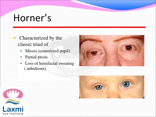
1. Horner’s syndrome:
2. Deep fascia of neck:

3. Structures in midline of neck:
Hyoid bone ⇒ Thyrohyoid membrane ⇒ Thyroid cartilage ⇒ Cricothyroid membrane ⇒ Cricoid cartilage ⇒ Cricotracheal ligament ⇒ Tracheal rings

NB: Internal laryngeal artery and superior laryngeal vessels pierces thyrohyoid membrane
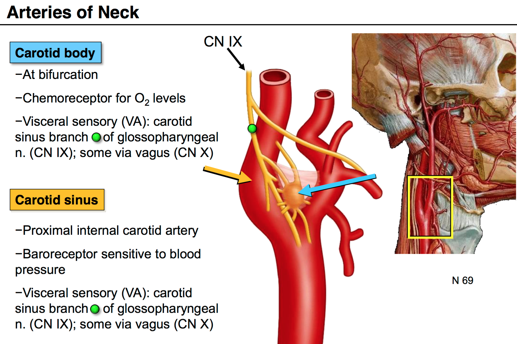
4. Carotid body and carotid sinus:
5. Parts of mandible:

6. External and internal auditory meatus:

EAM to tympanic cavity:
- Blood supply: Posterior auricular, superficial temporal
- Nerve: Great auricular, auriculotemporal
IAM:
- Blood supply: ascending pharyngeal
- Nerve: Glossopharyngeal
Clinicals:
- Otitis – ear inflammation
- Mastoiditis – middle ear infection
7. Fascial space infection:
These are summarized notes from various sources, mainly TeachMeAnatomy and Wikipedia
