Development of larynx, trachea, lungs and esophagus
Respiratory system
Origin: Endodermal floor of pharynx behind hypobranchial eminence
1. Epithelium
- Laryngo-tracheal groove appears in the endodermal floor of pharynx behind hypobranchial eminence
- Edges of groove unite dividing upper foregut into esophagus and laryngo-tracheal tube
- Laryngo- tracheal tube grows caudally:
- Upper end – larynx
- Then – trachea
- Lower part – 2 lung buds
- Right lung bud divides into 3 branches (main bronchii)
- Left lung bud divides into 2 bronchii
- Each bronchus – subdivisions – bronchial tree
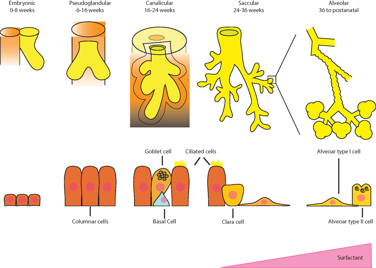
- Terminal/respiratory bronchioles end in alveoli, expand only after birth
2. Cartilage, muscles and connective tissue: 4th and 6th arch mesoderm
- 4th pharyngeal arch – thyroid cartilage, cricothyroid muscle
- 6th pharyngeal arch – cricothyroid, arytenoid, cuneiform and corniculate cartilages, and all other laryngeal muscles
NB: Splanchnic and somatic pleura forms lungs viceral and parietal pleura

Maturation of lungs:

Anomalies of lungs:
- Agenesis of one or both
- Accessory lung lobe
- Hyaline membrane disease – alveoli cannot ventilate adequately due to absence of surfactant
Esophagus
Origin: Endoderm of foregut
- From respiratory diverticulum to stomach swelling
- Surrounding mesenchyme forms musculature of esophagus
Anomalies of trachea and esophagus:
- Tracheal-esophageal fistula – incomplete fusion of laryngo-tracheal groove
- Esophageal atresia – narrowing of esophagus
- Failure of elongation – pulls stomach up thorax – Hiatus hernia
