Skeletal muscles
Origin: Paraxial mesoderm – myotomes
Myotomes ⇒ myoblast cells ⇒ fuse ⇒ myotubes ⇒ myofilaments develop in myotubes ⇒ myotubes become myocytes (muscle cells)
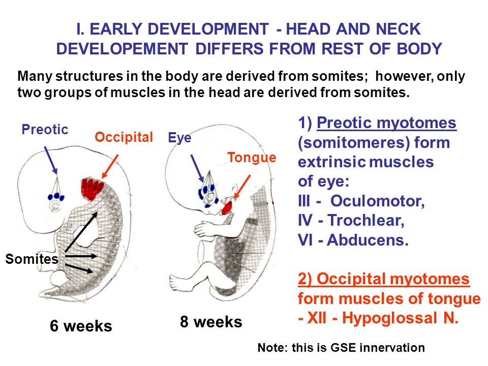
- 1st occipital myotome – extra occular muscles of the eye (NB: iris and ciliary muscle – neural ectoderm)
- Remaining 3 occipital myotomes – muscles of tongue
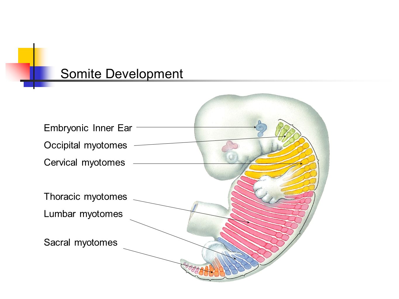
The 8 cervical, 12 thoracic, 5 lumbar, 5 sacral, 8-10 coccygeal myotomes divide into:
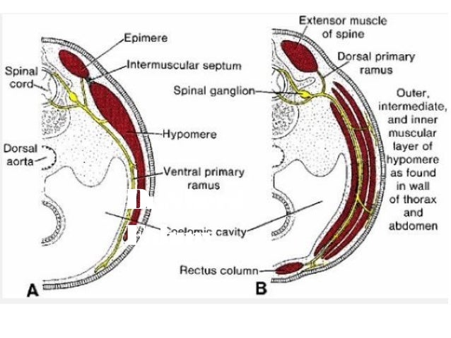
- Dorsal part – Epimere – forms extensor muscles of back and neck
- Ventral part – Hypomere:
- Split longitudinally eg. infrahyoid muscles
- Split to form layers eg. intercostal muscles
- Ventral tips unite to form eg. rectus abdominis muscle
NB:
- Myotomes migrate to limb buds to form flexor and extensor muscles
- Pharyngeal arches give muscles in head and neck
Limbs
- Upper limb bud forms opposite C5-T1
- Lower limb bud forms opposite L4-S3
Each limb bud is formed of:
- Lateral plate mesoderm – somatic layer – bones, tendons and connective tissue
- Migrating myotomes – muscles
- Neural crest cells – melanocytes and schwann cells
- Which is all covered by ectoderm – skin
Development:
- Programmed apoptosis between digital rays form fingers and toes
- Central mesoderm – cartilagenous skeleton – ossify into bone
- 3 segments formed in each limb bud
- Rotation of 90 degrees around long axis:
- Upper limb adducted, rotated laterally, thumb lateral
- Lower limb adducted, rotated medially, big toe is medial
Congenital anomalies of limbs:
- Amelia – failure to develop limbs
- Phocomelia – absence of proximal limbs, direct foot or hand
- Polydactyl – Extra fingers or toes
- Syndactyl – fusion of 2 fingers or toes
- Claw feet – lobster deformity
- Congenital digit amputation
- Constriction band
- Club foot
- Ectodactyl – missing middle finger
