All posts by DentMistry

History, clinical examination and treatment planning

History

Biodata

  1. Date
  2. File number
  3. Full name
  4. Age/date of birth
  5. Gender
  6. Contact
  7. Physical address
  8. Occupation
  9. Name of guardian/parent – for children
  10. Source of referral (if referred)

Presenting complaint

In patients own words, what is the problem

History of presenting complaint

  • S – Site
  • O – Onset
  • C – Character (throbbing, continuous, dull, acute, sharp)
  • R – Radiation (to head)
  • A – Associated symptoms (fever, discharge)
  • T – Timing (day or night, after eating)
  • E – Exacerbating factors (hot/cold food), Alleviating factors (Pain medications)
  • S – Scale (scale of 1-10, rate the pain)

Previous treatments concerning the presenting complaint

Past dental history

  • Index visit or
  • Previous dental treatments done
    • What they were
    • When
    • Where
    • If extraction done – any complications
    • Tolerance to LA

Dental habits

  • How many times do they brush their teeth
  • How do they brush
  • Which toothpaste
  • How often they change their brush
  • Any interdental cleaning methods used – floss, toothpicks
  • Abnormal habits eg. mouth breathing, lip sucking

Past medical history

  • History of chronic illness:
    • CHD/CVS, infective endocarditis
    • Respiratory – asthma, bronchitis
    • GIT – peptic ulcers, diarrhea, vomiting, jaundice, hepatitis, gastritis
    • Diabetes
    • CNS disorders
    • Bleeding disorders – hemophilia, anticoagulant therapy
    • Infectious diseases – TB, HIV, Herpes
    • On any medications – NSAID, corticosteroids, anticoagulants, anticonvulsants
  • Previous hospital admission – When, where, why, treatment provided
  • Food or drug allergy

Obs and gyn history for females

  • Last menstruation date and regularity
  • Pregnancy status
  • Type of contraceptives used

Family social history

  • Alcohol – amount and frequency
  • Smoking – amount and frequency
  • Drugs
  • Family status – parents, siblings, chronic illness in family
  • Martial status and children
  • Water source – borehole or city council

For pediatric and orthopedic patients

Birth history:

Prenatal:

  • Health and nutritional status of mother during pregnancy
  • Complications during pregnancy:
    • Infections – rubella, TB, syphilis, UTI
    • Pre-eclampsia
    • Hypertension
    • Diabetes
    • Antepartum bleeding
  • Drugs
  • X-ray
  • Rh incompatibility may result in erythroblastosis fetalis – leading to green blue discoloration of dentition. Picture

Natal:

  • Full term or premature
  • Mode of delivery – Normal/C-section and why?
  • Did the baby cry on birth
  • Birth weight
  • Breast fed or formula milk given

Postnatal:

  • Vaccinated
  • Developmental history
  • Nocturnal feedings/sweetened milk – predisposes to early childhood caries (read more)
  • Brushing habits – frequency, by who, supervised?

Habits

  • Finger sucking/thumb sucking
  • tongue thrusting
  • Mouth breathing
  • Nail biting – check nails

Diet chart

  • 24 hour diet chart
  • 7 day diet chart (as investigation)

Family social history

  • Name of school
  • Class
  • Performance in school
  • Social or antisocial
  • Occupation of parents
  • Family history
  • Water source

Clinical examination

General examination

  • Anxious or calm
  • Build, nourishment – well, poor
  • Posture

Vital signs:

  • Temperature
  • Pulse rate
  • Respiratory rate
  • Blood pressure

NB: Also measure weight and height for children – to calculate BMI and dosage of LA and drugs

Extra oral examination

  • Palpate submental, submandibular and neck lymph nodes
  • Facial symmetry – any swellings or asymmetry
  • Facial profile
  • Scars
  • Eyes – jaundice (look down), pallor (look up)
  • TMJ movements – clicking or popping sounds, pain, path of closure
  • Lips competency
  • Hands – examine nails, finger clubbing, cyanosis

Remember it as: J A C C L O W D (Jaundice, anemia, clubbing, cyanosis, lymphadenopathy, oedema, wasting, dehydration)

Intraoral examination

  • Oral hygiene status
  • Type of dentition: primary, mixed, permanent

Soft tissue examination:

  • Gingiva – shape, size, color, bleeding, ulceration, growths, pockets, recession
    • Plaque and gingival score
  • Buccal mucosa – color, texture, ulcer, growth, sinus
  • Floor of mouth – swellings, ulcer
  • Tongue – size, movements, plaque
  • Palate – normal, high vault, clefts
  • Tonsils – normal, swollen
  • Frenal attachments – normal, higher

Hard tissue examination:

  • According to quadrants
  • Teeth present
  • Teeth missing
  • DMF
  • Palpate, percuss
  • Check interproximal caries with floss
  • Wear (attrition, abrasion, erosion)
  • Discoloration
  • Malformation
  • Mobility – Millers classification 1950
    • 0 = No detectable mobility
    • 1 = Distinguishable mobility
    • 2 = Horizontal movement > 1mm
    • 3 = Horizontal and vertical movement > 1mm
  • Orthodontic assessment
  • Fluorosis – TF score for every tooth
TF score for fluorosis

Tooth fracture classification

Periodontal assessment

Gingiva:

  • Color: Pink, physiologic pigmentation, red, cyanotic
  • Size: Mild, moderate, severe inflammed
  • Shape: Scalloped, rounded, col – if space between 2 teeth
  • Consistency: Firm, flabby
  • Texture: Stippling on attached gingiva

Oral hygiene:

  • Calculus – presence of supra or subgingival calculus
  • Plaque seen with naked eye

Gingival index by Loe and Silness 1963:

  • Facial and lingual surface of index teeth: 16, 11, 24 and 36, 31, 44
  • Rate:
    • 0 = Normal
    • 1 = minimal inflammation, erythema, no bleeding
    • 2 = Bleed on probing
    • 3 = Spontaneous bleeding
  • Find mean score
  • GI score:
    • 0-1 = Mild
    • 1-2 = Moderate
    • 2-3 = Severe

Periodontal index by Turesky et al modified Quigley Hein 1970:

Index teeth and disclosing tablet

  • 0 = No plaque
  • 1 = Flecks at cervical margin
  • 2 = Thin continuous band at cervical margin
  • 3 = Band wider than 1mm, < 1/3 of crown
  • 4 = Plaque < 2/3 of crown
  • 5 = Plaque > 2/3 of crown

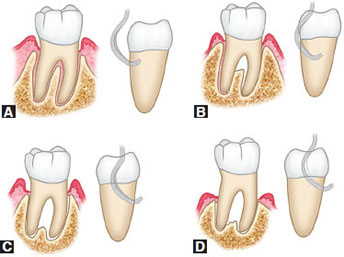
Furcation involvement – Glickman classification 1953

  • Grade 1 – Incipient, pocket formation into furcation fluting, interradicular bone intact
  • Grade 2 – Moderate loss of interradicular bone but not through and through
  • Grade 3 – Probe goes through and through, orifice occluded by gingival tissue
  • Grade 4 – Exposed furcation

Periodontal charting:

  • Draw continuous line of free gingival margin – facial and lingual
  • Draw interrupted line indicating bone level on facial side
  • Record 6 point pocket depth
  • Record tooth mobility
  • Record missing teeth (X), open contacts(//) and how many mm, overhang restorations (V)
  • Calculate clinical attachment loss (CAL): Gingival recession + pocket depth
    • 1-2mm = Mild
    • 3-4mm = Moderate
    • > 5mm = Severe
    • Localized: < 30% of sites
    • Generalized: > 30% of sites

Orthodontic assessment

  • A-P: Molar, canine, incisor relationship – Class I, II, III
  • Vertical: Open bite, overbite – deep or open
  • Transverse:
    • Crossbite anterior or posterior
    • Midlines
    • Crowding or spacing
    • Rotation or displacement
    • Proclination
  • Upper and lower arch form – Normal, V shape, square shape

Investigations

  • Radiological – describing a x-ray:
    • Name and age of patient
    • Date when the x-ray was taken
    • Quality
    • Teeth present
    • Dental age and why
    • Radiolucent/radiopaque lesions
  • Microbiological
  • Histopathological
  • Study model
  • Diet chart
  • Plaque score
  • BMI

Diagnosis

  • Summarize findings: eg. A 5 year old African male with early childhood caries, dentoalveolar abscess secondary to extensive decay on 55, irreversible pulpitis 85 and 75, and occlusal caries on 54, 64, 84, 74
  • For periodontal diagnosis: Severity – extent – diagnosis, eg:
    • Mild – localized – chronic periodontitis
    • Moderate – generalized – plaque induced gingivitis secondary to orthodontic treatment and mouth breathing

Prognosis for periodontology

1. Excellent prognosis:

  • No bone loss
  • Excellent gingival condition
  • Good patient co-operation
  • No systemic/environmental factors

2. Good prognosis:

  • Adequate remaining bone support
  • Adequate control of etiologic factors and maintainable dentition
  • Adequate patient co-operation
  • No systemic/environmental factors or well controlled

3. Fair prognosis:

  • Less than adequate bone support
  • Some tooth mobility
  • Grade 1 furcation
  • Adequate maintenance possible
  • Acceptable patient co-operation
  • Presence of limited systemic/environmental factors

4. Poor prognosis:

  • Moderate-advanced bone loss
  • Tooth mobility
  • Grade 1 or 2 furcation involvement
  • Doubtful patient co-operation
  • Presence of systemic/environmental factors

5. Questionable prognosis:

  • Advanced bone loss
  • Tooth mobility
  • Grade 2 or 3 furcation involvement
  • Inaccessible areas
  • Systemic/environmental factors

6. Hopeless prognosis:

  • Advanced bone loss
  • Extraction indicated
  • Non maintainable areas
  • Uncontrolled systemic/environmental factors

NB: Factors affecting prognosis:

  • Diagnosis: Disease severity, plaque and calculus
  • Systemic factors: DM, puberty, genetic
  • Occlusal factors
  • Prosthetic and restorative factors: Caries, teeth vitality, abutment selection, subgingival restorations, fixed or removable prosthesis
  • Patient factors: Compliance, co-operation, attitude
  • Environmental factors: Smoking, alcohol. bruxism

Treatment objectives

  1. To control infection and relieve pain and discomfort
  2. To modify attitude to dental care, and behavior to dental treatment
  3. To improve oral hygiene
  4. To restore integrity and function of the dentition
  5. To achieve cariostasis
  6. To improve esthetics and correct malocclusion
  7. To maintain a healthy oral cavity
  8. Diet planning

Treatment planning

  1. Oral hygiene instruction (OHI)
  2. Emergency phase: Systemic diseases, infections
  3. Etiological phase: Plaque and calculus
  4. Restorative phase: Filling, RCT, Prosthetic replacement
  5. Maintenance phase: Recall and review

Periodontal treatment planning

  1. Preliminary phase: Systemic disease, infections, OHI
  2. Etiological phase: FMS, root planing, fluoride treatment, cavity prep and filling
  3. Surgical phase: Disimpaction, gingivectomy, implants, open flap debridement, GTR, furcationplasty
  4. Restorative phase: Crown, bridge, crown for implant
  5. Supportive periodontal therapy/maintenance phase: Review after 2 weeks
    • High risk – recall every 3 months
    • Moderate risk – recall every 6 months
    • Low risk – recall every year

Pediatric treatment planning

  1. Systemic phase – Stabilize chronic illness before dental treatment
  2. Emergency phase – Antimicrobials
  3. Preventive phase – OHI, behavior management, fluoride application, diet counselling, pit and fissure sealants
  4. Preparatory phase – Oral prophylaxis, caries control if multiple lesions, preventive orthodontic consultation
  5. Corrective phase – Restorations, prosthetic replacement, extractions, Interceptive orthodontic consultation
  6. Maintenance phase – Recall and review, 3-6 months
  • Significance of oral prophylaxis:
    • Introduction to dental environment/behavior management
    • Oral hygiene education
    • Uncover carious lesion covered in plaque
    • Healthy gingiva

Orthodontic History, Examination, Investigation, Treatment Planning and Cephalometric Analysis

History:

  • Biodata
  • Presenting complain
  • HPC
  • PDH
  • Dental habits
  • PMH
  • FSH
  • Birth history
  • Abnormal habits

Clinical examination:

General examination

Extraoral examination:

  • JACCLOWD
  • TMJ movements – clicking or popping sounds, pain, path of closure
  • Facial profile
  • Facial symmetry
  • Lip competency
  • Incisor showing on smiling – Normally 2-4mm
  • Nasolabial angle and lip protrusion:
    • Between upper lip and base of nose
    • Normal 90° – 110°
    • Convex – Class II, retrusive mandible
    • Concave – Class III, protrusive mandible
    • Becomes retrusive and obtuse angle with age
Nasolabial angle
  • Vertical facial relationship – Angle of lower border of mandible to cranium
    • Use occipital region to draw imaginary line
    • Average
    • Reduced – Short face syndrome: Deep bite, overlapped lips
    • Increased – Long face syndrome: Anterior open bite, incompetent lips
Vertical facial relationship

Intraoral examination:

  • Oral hygiene status
  • Type of dentition: primary, mixed, permanent
  • Soft tissue and hard tissue examination
  • Dental arches:
    • Crowding: mild, moderate, severe
    • Spacing
    • Tooth rotations: mesioversion, distoversion lingoversion, buccoversion, labioversion)
    • Tooth displacement
    • Ugly duckling stage

Orthodontic examination:

  • Anterior – posterior plane: Molar, canine, incisor relationship – Class I, II, III
    • Molar malocclusions: Maxillary 6 MB cusp and mandibular 6 buccal groove.
      • Angles class I: 7 types:
        • 1. Maxillary teeth crowded
        • 2. Anterior teeth proclined
        • 3. Anterior crossbite
        • 4. Posterior crossbite
        • 5. Permanent molars drifted mesially
        • 6. Diastema
        • 7. Deep overbite
      • Angles class II:
        • Division I: Incisors proclined
        • Division II: Incisors retroclined
        • Subdivision: if right or left unilateral Angles class II
      • Angles class III:
        • Type 1: Edge to edge bite
        • Type 2: Normal overbite
        • Type 3: Anterior cross bite
        • Pseudo class III malocclusion: Mandible moves forward
  • Vertical plane:
    • Overbite: Normal 20-40%. <20% = reduced overbite, >40% = deep bite
    • Open bite
    • Anterior crossbite/ reverse overjet
  • Transverse plane:
    • Midline
    • Crossbite
    • Scissor bite

Investigation

  • OPG, study model, photograph, lateral cephalogram, CT scan

OPG:

  • Name and age of patient
  • Date when the x-ray was taken
  • Quality
  • Teeth present
  • Dental age, development of crown and roots, root completion 2-3 years after eruption
  • Radiolucent/radiopaque lesions

Study model analysis: Note date of impression, patient name, D.O.B, file number

a) Interarch analysis:

  • A-P, transverse, vertical plane
  • Use dividers to measure

b) Intra arch analysis: Maxillary and mandibular

  • Shape (U, V)
  • Arch symmetry (position of teeth, missing teeth)
  • Palate vault
  • Number of teeth present, or eupting
  • Individual tooth malformation, malposition, or rotation

c) Space analysis:

  • Arch parameter (X) = Measure arch from 5 to 5, distal surface
  • Tooth material (Y)= Width of each tooth. Angles lines of occlusion:
    • Maxillary – use central fossa and cingulum (in anterior teeth)
    • Mandible – use buccal cusp tips and incisal edges
  • Difference between arch parameter and tooth material:
    • X – Y = Positive (spacing), Negative (crowding)
    • ≤ 4mm = Mild crowding
    • 5-8 mm = Moderate crowding
    • > 9mm = Severe crowding

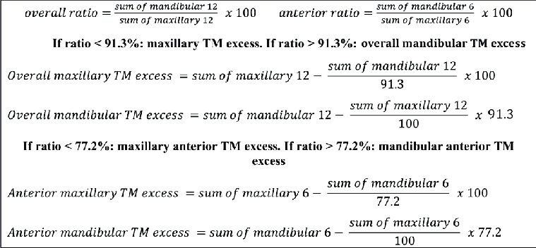
d) Bolton’s analysis: Maxillary and mandibular relationship for overbite/overjet

  • Sum of mesiodistal width of 12 teeth: CI, LI, C, PM, PM, M1 on both sides

Sum of mandibular 12/Sum of maxillary 12 X 100 = 91.3% ± 1.91 (ie. range: 89.39-93.21)

  • < 91.3% = Maxillary teeth in excess
  • >91.3% = Mandibular teeth in excess

e) Anterior ratio: Maxillary and mandibular relationship for overbite/overjet

  • Sum of mesiodistal width of 6 teeth: CI, LI, C on both sides

Sum of mandibular 6/Sum of maxillary 6 X 100 = 77.2% ± 1.65 (ie. range: 75.55-78.85)

  • < 77.2% = Anterior maxillary teeth in excess
  • >77.2% = Anterior mandibular teeth in excess

NB: Bolton’s analysis and anterior ratio cannot work if required teeth are missing

Bolton's analysis and anterior ratio

Photographs: Smile and profile analysis, record keeping

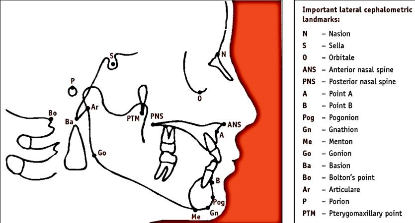
Lateral cephalogram analysis

Mixed dentition analysis using study models:

a) Radiograph/Huckaba analysis:

True width of 1st molar/Apparent width of 1st molar = True width of unerupted PM/Apparent width of unerupted PM

b) Moyer’s prediction table:

  • Use sum of mandibular 4 incisors to predict mesiodistal width of permanent canine and premolars
  • 75 percentile usually used
Moyer's prediction table

c) Tanaka and Johnston:

  • Estimated mesiodistal width of canine and premolar of one quadrant = 1/2 of the mesiodistal width of mandibular 4 incisors + 10.5mm (for mandible) or 11mm (for maxilla)

d) Nance – Arch perimeter analysis:
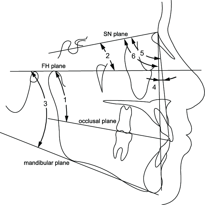
  • Mesiodistal width of erupted permanent teeth and from IOPA of unerupted teeth

Diagnosis

  • List name, age and gender
  • Write problem list in priority
  • Eg. Angles class I malocclusion with an anterior open bite extending from 15/45 to 25/35, with an overjet of 6mm and tongue thrusting habit

Treatment plan

  • List treatment objectives according to PC and priority
  • List treatment plan

Cephalometric analysis

Cephalometric landmarks

  • S – Sella turcica – center of pituitary fossa
  • N – Most anterior point of frontal and nasal bone junction
  • A – Inner most point between ANS and incisor
  • B – Inner most point between mandible and incisor
  • Pog – Anterior most point of mandible
  • Gn – Most anterior inferior point of bony chin
  • Go – Point where posterior border of ramus and lower border of mandible bisect
  • Porion – External auditory meatus upper contour midpoint
  • Orbitale – Inferior margin of orbit – lowest point
  • Frankfort plane (FH) – Porion to orbitale
  • Occlusal plane – Use molars and premolars
  • Mandibular plane – Gn to Go
Cephalometric planes

Steiner’s analysis

Skeletal analysis:

1. SNA angle: 82° ± 2

  • Ant-post position of maxilla with cranial base
  • Increased angle = Prognathic maxilla

2. SNB angle: 80° ± 2

  • Ant-post position of mandible with cranial base

3. Angle ANB: 3 ± 1

  • Difference between SNA and SNB – magnitude of skeletal jaw discrepancy
  • Factors affecting:
    • Vertical height of face
    • Abnormal position of nasion
  • Increased angle = Class II
  • Decreased angle = Class III

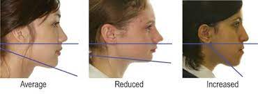
4. Mandibular plane angle: 32° ± 4

  • Steepness of mandibular plane to cranial base
  • Increased angle = Vertical growth
  • Decreased angle = Horizontal growth

5. Occlusal plane angle: 17° ± 4

  • Determine relationship of teeth in occlusion with cranial base
  • Increased angle = Skeletal open bite
  • Decreased angle = Skeletal deep bite

Dental analysis:

1. UI – NA angle and distance: 22°, 4mm

  • > 4mm or increased angle = Protrusion eg. class II division 1
  • < 4mm or decreased angle = Retrusion eg. class II division 2

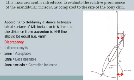
2. LI – NB angle and distance: 25°, 4mm

  • > 4mm or increased angle = Protrusion eg. class II division 1
  • < 4mm or decreased angle = Retrusion eg. class II division 2/ class III

3. Interincisal angle: 130°-131°

  • Increased angle = Class II division 2
  • Decreased angle = Class II division 1

4. Lower incisor to chin (Holdaway ratio)

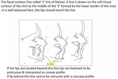
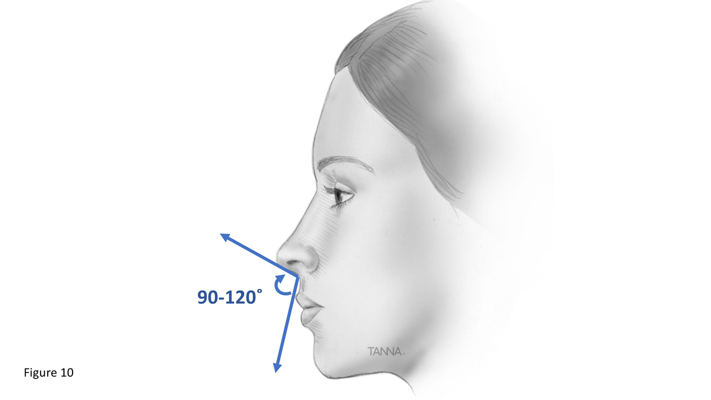
Soft tissue analysis:

Langerhans cell histiocytosis

Formerly known as histiocytosis x (eosinophilic granuloma, hand-schuller-christian disease, letterer-siwe disease)

  • Due to proliferation of abnormal histiocytes (subtypes of WBC)

Clinical: Short stature, diabetes insipidus, neurosensory deafness and tooth mobility

Radiology: Osteolysis causing ’floating teeth’ in multiple quadrants

Histology:

  • Diffuse infiltration of large, pale staining mononuclear cells with indistinct cytoplasmic
    borders rounded/bean shaped nuclei
  • Birbeck granules: rod shaped cytoplasmic structures characteristic to Langerhans cells (s100 positive)

Management: Curettage of accessible bone lesions

Picture

Scurvy

  • ↓ Ascorbic acid (vit C)

Physiology: Vit C is required for redox reactions:

  • Hydroxylation of praline in collagen formation
  • Hydroxylation of dopamine to noradrenaline

Clinical: Scurvy

  • Haemorrhagic diatheses
  • Skin rash
  • Delayed wound healing
  • Anaemia
  • Scorbutic rosary – mineralization of costochondral cartilages due to deranged formation
    of osteoid
  • Generalized gingival enlargement

Histology: Hemorrhagic gingival enlargement with minimal fibrosis

Management: Replacement therapy

Picture

Renal osteodystrophy and rickets

  • Skeletal abnormalities due to chronic renal failure
  • Kidneys are vital of vit D metabolism, and phosphate secretion – when disturbed results in:
    • Osteomalacia (or rickets)
    • Secondary hyperparathyroidism
    • Metastatic calcification
    • Osteoporosis
Chronic renal failure flow chart

Rickets:

Deficiency of vitamin D during bone development (infancy)

  • Essential for absorption and metabolism of calcium and phosphate
  • Deficiency leads to:
    • Defect in bone matrix mineralization and skeletal development
    • Defective absorption of calcium and phosphate
    • Chronic renal disease: renal rickets

Etiology:

  • 1. Vitamin D deficiency
    • Reduced endogenous synthesis due to lack of exposure to the sun
    • Dietary deficiency (fish, eggs, butter, milk)
    • Malabsorption and metabolism failures due to liver disease, pancreatic insufficiency, kidney
  • 2. Abnormal metabolism of vitamin D:
    • Chronic renal failure
    • Vitamin D resistant rickets
    • Anticonvulsant osteopathy
  • 3. Phosphate depletion: antacids contain al(oh)3 which bind phosphate, excess renal secretion of po4
    • Fanconi syndrome
    • X linked hypophosphatemic rickets
  • 4. Renal tubular acidosis

Physiology:

  • Vit D is essential for intestinal absorption of Ca++ and po4
  • Co-factor in mobilization of Ca++ from bone
  • Stimulates PTH dependent re-absorption of Ca++ by the distal renal tubules

Clinical:

  • Craniotabes – unossified areas of cranium
  • Rachitic rosary – overgrowth of costochondral cartilage
  • Pigeon chest deformity
  • Lordosis
  • Knock knees/ bow legs
  • Enlarged epiphyses
  • Osteomalacia
  • Hypocalcaemic tetany
  • Dental:
    • Multiple spontaneous periapical abscess in 1ry & 2ry dentition
    • Enlarged pulp chambers
    • Thin easily abraded enamel
    • Marked interglobular dentine

Histology: ↑ Osteoid matrix in bones

Lab:

  • Normal or ↓ ca++
  • ↓ po4
  • ↓ vit D metabolites
  • ↑ alkaline phosphatase

Management: Replacement therapy

Picture

Osteoporosis

  • Quantitative reduction of normal bone → risk of fractures, pain, bone deformity

Etiology/classification:

1. Primary – Osteopenia without an underlying disease or medication

  • Idiopathic type – rarely, in young pt
  • Involutional type – postmenopausal women and geriatric patient

Contributing factors:

  • Race (more in caucasians and asians)
  • Gender: F > M.
  • Reduced physical activity (convalescence and old age)
  • Oestrogen, calcitonin and androgen deficiencies
  • Malnutrition states
  • Hyperparathyroidism
  • Vitamin d deficiency

2. Secondary – due to:

A. Endrocrine disorders:

B. Neoplasia: multiple myeloma, carcinomatosis
C. Git problems: malnutrition, malabsorption, hepatic insufficiency, vit c & d deficiency
D. Medication: corticosteroids, anticonvulsants, heparin, alcohol
E. Miscellaneous: immobilization, anemias, pulmonary disease

Radiology: Enlargement of the medullary cavity + thinning of cortex

Histology:

  • Active type – ↑ bone turnover – by osteoblastic and osteoclastic activity with new osteoid formation
  • Inactive osteoporosis – almost normal bone structure

Lab: ↑ serum phosphatase

Management: Hormone replacement therapy, manage other underlying causes

Picture

Osteitis deformans – Paget’s disease

Abnormal and anarchic resorption and deposition, resulting in distortion and weakening of the affected bones

Epidemiology: M > F, patients over 50 years, Max > Mand (2:1)

Etiology:

  • Slow paramyxovirus infection
  • Vascular disorder
  • AD

Pathology:

  • Fast, irregular, exaggerated bone remodelling
  • Thick bones without localized swelling
  • Early phase – resorption
  • Late stage – sclerosis
  • Patchy appearance due to areas of resorption and sclerosis adjacent to each other

Clinical:

  • Simian stance due to weakening of weight bearing bones,
  • Lion-like facial deformity (bilat symmetrical bone enlargement)
  • Pain on involved bones
  • Hypercementosis
  • Neurological symptoms due to narrowing of foramina:
    − Headache
    − Vertigo
    − Visual disturbance
    − Auditory disturbance

Complication: (0.9 – 13%): progression to osteosarcoma

Histology:

  • Initial osteolytic – osteoclastic bone resorption
  • Mixed osteolytic/osteoblastic stage -mosaic pattern of reversal lines (osteoid seams)
  • Quiescent osteosclerotic stage – dense bone with remnants of mosaic patterns

X-ray:

  • “Cotton wool” appearance of confluent radio-opacities
  • Thickening of bone
  • Irregular areas of sclerosis and resorption
  • Loss of normal trabeculation

Lab:

  • ↑alkaline phosphatase
  • Normal calcium and phosphorus levels
  • ↑ urinary hydroxyproline

Management: Seldom fatal

  • Surgical debulking + calcitonin (PTH antagonist) + sodium phosphatase (retards bone resorption)

Picture

Hypothyroidism

  • ↓ Thyroid hormone

Etiology:

  • Congenital defects of the thyroid
  • Iodine deficient goiter
  • Autoimmune (Hashimoto’s) thyroiditis
  • Diseases of the pituitary (↓TSH)
  • Hypothalamus (↓ thyrotropin releasing hormone TRH)
  • Radiation injuries
  • Surgical ablation
  • Drugs (lithium, iodides)
  • Idiopathic causes

Clinical:

  • Delayed mental, skeletal, dental and sexual development
  • Blood: microcytic, hypochromic anemia – fatigue and lethargy
  • Dermal: dry and scaly skin, facial oedema.

Management: Gradual replacement therapy + synthetic and natural thyroid hormone preparations.

Picture