Category Archives: Oral Pathology

Pemphigus and Pemphigoid

Pemphigus vulgarisMucous membrane pemphigoid
Etiology
Target proteins
– Autoimmune reaction
– Intercellular keratinocyte protein (desmoglein 3) destroyed by auto antibodies
– Autoimmune reaction to basement membrane proteins:
– Lamina 5
– BP 180
ClinicalIntraepithelial blistersSubepithelial blisters
Site– Oral mucosa
– Skin
– Mucosa
– Oral mucosa
– Conjunctiva
– Skin rarely
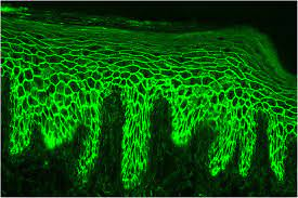
Histology– Intraepithelial clefting
– Loss of desmosomal contacts therefore free floating acantholytic Tzanck cells
– Subepithelial clefting
– Chronic inflammatory infiltrate
Direct immunofluorescence testingCirculating auto IgG No circulating auto IgG
Difference between pemphigus and pemphigoid

Pemphigus vulgaris

  • Autoimmune blistering disorder
  • Results in loss of integrity of intercellular attachments within epidermis and mucosal epithelium

Subtypes:

  • P. Vulgaris (common)
  • P. Vegetans (wart like)
  • P. Foliaceus (flower like)
  • P. Erythematosus (reddened)

Nikolsky’s sign: Positive – stroke mucosa gently, vesicle or bulla appears

Management: Corticosteroids + Azathioprine + Cyclophosphamide

Prognosis: Good. Fatal if untreated

Immunofluorescence of pemphigus vulgaris

Mucous membrane pemphigoid

  • Autoimmune blistering disorder
  • Results in loss of integrity of dermal-epidermal junctions
  • Affects oral tissues in 30% of cases
  • More severe

Management: Corticosteroids

Prognosis: Fair

Differential diagnosis:

  • Erythematous lichen planus
  • Linear IgA disease
  • Discoid lupus erythematous
  • Contact allergy
immunofluorescence of mucous membrane pemphigoid

Aphthous ulcer

Etiology

  • Immunological disorder – T cell mediated
  • Neurogenic inflammation
  • Nutritional deficiency – B12, folic acid, iron
  • Microbial – Viral, bacteria
  • Mucosal healing defects – by cytokine inhibition

Clinical

  • Burning/tingling sensation
  • Painful recurrent ulceration
  • Ulcer on non keratinized mucosa
    • Slightly raised erythematous margin
    • Necrotic base

Histology

  • Non specific inflammation + macrophages and mast cells
  • Absence of viral infected cells
  • Differential diagnosis: Herpetic gingivostomatitis

Subtypes

Minor aphthaeMajor aphthaeHerpetiform aphthae
Size0-0.5cm>0.5cm0-0.5cm
ShapeOvalRagged oval, crateriform Oval
Number1-101-510-100
LocationNon keratinized mucosaNon keratinized mucosaAny intra oral site
Subtypes of aphthous ulcer

Management

  • Corticosteroid therapy + tetracycline mouthwash

Picture

HIV/AIDS

Difference between HIV and AIDS

Etiology

  • RNA retrovirus – Human Immunodeficiency Virus

Pathology

  • HIV – specific affinity for – CD4+ T helper cells – Therefore damage cell mediated immune system
  • Enters host – replicates using host DNA – stay latent – cell fusion/cell death
  • Notes under Virology

Modes of spread

  • Vertical transmission and breast feeding
  • Sexual contact
  • Parental exposure:
    • Organ transplant
    • Blood transfusion
    • Contaminated needles
    • Artificial insemination

Clinical

1. Systemic signs and symptoms

  • Chronic fever
  • Weight loss
  • Diarrhea
  • TB
  • Persistent generalized lymphadenopathy
  • Encephalopathy (AIDs dementia)
  • Other STIs

2. Oral infections

  • Fungal:
    • Candidiasis
    • Histoplasmosis
    • Cryptococcus neoformans
    • Penicilliosis
  • Protozoa: Leishmania

3. Oral neoplasms

4. Other manifestations

Lesions commonly and least commonly

Diagnosis

  • ELISA
  • PCR
  • CD4 counts
  • CD4 : CD8
  • Indirect immunofluorescence – HIV specific IgM

Management

  • ARVs eg. Zidovudine, Ritonavir

Clinical features of pediatric AIDs

Age of onset: Neonate to late infancy

Infections:

  • Fungi – C. albicans
  • Mycobacteria – M. TB, M. bovis, atypical
  • Bacteria – S. pneumonia, S. aureus, Gram -ve’s
  • Virus – HSV, EBV, VZV, CMV, adenovirus
  • Protozoa – P. carinii (Update: Now considered a fungus by the name Pneumocystis jirovecii)

Malignancy:

Nutrition:

  • Diarrhea
  • Failure to thrive

Systemic:

  • Fever
  • Lymphadenopathy
  • Hepatosplenomegaly

Parotid: Diffuse, non suppurative enlargement

CNS: Developmental delays

AIDS dysmorphic syndrome: Variable teratogenicity

Actinomyces

Etiology

  • Actinomyces israelii

Subtypes

  • Cervicofacial
  • Abdominal
  • Pulmonary
  • Cutaneous

Predisposing factors

  • Surgery and dental procedures
  • Previous infection
  • Osteoradionecrosis
  • Trauma
  • Systemic illness

Mnemonic: SPOTS

Clinical

  • Indurated woody hard swelling + multiple discharging sinuses Picture
  • Pus discharge – Yellow and granular (sulfur granules)
  • Chronic low grade persistent infection

Histology

  • Sulfur granules
  • Filamentous rays in round colonies surrounded by pleomorphs, giant cells and macrophages

Management

  • Penicillin + surgical debulking

Tuberculosis

Etiology

  • Mycobacterium tuberculosis
  • Oral lesion secondary to pulmonary TB
  • AIDS defining illness

Clinical

  • Scrofula – lymphadenopathy on neck Picture
  • Fever
  • Night sweats
  • Malaise
  • Weight loss
  • Cough
  • Hemoptysis
  • Intraoral lesion: Indurated chronic non healing ulcer and is painful

Diagnosis

NB: Immunocompromised patients have atypical mycobacteria infections eg. M. avium intracellulare

Histology

  • Caseating necrosis/granuloma – cheesy appearance
  • Langerhans giant cells
  • Inflammatory response

Management

  • Directly observed therapy (DOT) – multidrug therapy
  • Isoniazid
  • Rifampicin
  • Ethambutol
  • Pyrazinamide
  • Streptomycin

Mnemonic: PRIESt

Clinical stages

  • Non immune host exposed to TB (skin test positive)
  • Primary TB of lung
  • Arrested TB in immunocompetent individuals
  • Progressive TB – hematogenous, lymphatic, direct spread
  • Arrested TB reactivated – secondary TB – progressive TB

Cervical TB:

  • Most common in level 2 nodes
  • Lymph nodes > 2cm
  • With or without evidence of TB elsewhere
  • Clinical: Night sweats, weight loss, fever
  • Diagnosis: FNA, ZN staining with AFB, Mantoux test

Syphilis (treponema pallidum)

1. Sexually transmitted (acquired)

Primary – Chancre (chronic ulcer) Picture

Secondary

Tertiary

  • Gumma (non cancerous growth) Picture
  • Destructive ulcers
  • CNS disease – Neurosyphilis:
  • CVS disease – Aortitis:
    • Aneurysms
    • Aortic regurgitation and gumma

NB: Can go into latency


2. Vertically transmitted (congenital)

Early

  • Spirochetosis
  • Dental defects
  • Deafness
  • Ocular Keratitis
  • Bone and joint lesion
  • Organopathy

Late: Latency


Diagnosis

Management

  • Penicillin
  • Tetracycline

Notes under white lesions

Prenatal infection:

  • Hutchinson’s incisors – tapered crown + notched incisal edge
  • Mulberry molars – Lobulated occlusal surfaces

Picture

Ulcerative lesions

Definitions

Primary and secondary skin lesions

1. Macule

  • Focal area of colour change
  • No elevation or depression in relation to surroundings

2. Papule

  • Solid raised lesion
  • Less than 5mm in diameter

3. Nodule

  • Solid raised lesion
  • More than 5mm in diameter

4. Sessile

  • Tumor/growth
  • Base is widest part of lesion

5. Pedunculated

  • Tumor/growth
  • Base is narrower than widest part of lesion

6. Papillary

  • Tumor/growth
  • Numerous warty projections

7. Verrucous

  • Tumor/growth
  • Rough warty surface

8. Vesicle

  • Superficial blister filled with clear fluid
  • Less than 5mm in diameter

9. Bulla – Blister > 5mm in diameter

10. Pustule – Blister filled with purulent exudate

11. Ulcer

  • Loss of surface epithelium ± underlying CT
  • Depressed/ excavated

12. Erosion

  • Superficial lesion
  • Partial/ total loss of surface epithelium
  • After rupture of vesicle or bulla

13. Fissure – Narrow slit like ulceration/ groove

14. Plaque – Elevated and flat on surface

15. Petechia – Round pinpoint area of hemorrhage

16. Ecchymosis – Non elevated hemorrhage, larger than petechia

17. Telangiectasia

  • Vascular lesion
  • Dilation of small superficial blood vessel

18. Cysts

  • Pathological epithelium lined cavity
  • Filled with liquid or semisolid content

19. Unilocular – Radiolucent lesion – single compartment

20. Multilocular – Radiolucent lesion – several compartments


Traumatic lesions

Clinical manifestations of trauma

  1. Acute/chronic ulcers
  2. Red/white lesions
  3. Mucositis
  4. Reactive hyperplasia
  5. Bone exposure and sequestration

Etiology

Physical trauma

  • Factitial injury – Self induced/psychological
    • Eg. Cheek biting – subconscious reaction to stress, emotions, boredom
  • Riga-Fede disease – Traumatic ulcer on anterior tongue with natal teeth in infants, or repetitive tongue thrusting habit after eruption of 1ry lower incisors
  • Frictional hyperkeratosis – white lesion, ill fitting dentures
  • Iatrogenic

Chemical burns, thermal burns

Therapeutic radiation

Classification

1. Acute traumatic ulcer

  • Pain
  • Yellow base, red halo
  • Heals in 7 to 10 days

2. Chronic traumatic lesions

  • Little or no pain
  • Scar formation
  • Mimic carcinoma/infective ulcer

Clinical

  • Single lesion
  • Erythematous
  • Non inverted margins
  • Clean base covered in pseudomembrane
  • Painful
  • Disappear in 7-10 days after eliminating cause

Histology

  • Loss of epithelium – replaced by fibrin network
  • Granulation tissue base
  • Scar formation deep in tissues
  • Dense inflammatory infiltrate – macrophages and eosinophils

Management

  • Observe for 2 weeks to rule out infection
  • Topical corticosteroids

Pathological lesions

Etiology

1. Neoplasm

2. Immunological disease

3. Aphthous ulcer/ Canker sore – Multifactorial

4. Infections

Nb: Chronic infectious ulcers (TB, syphilis, fungal)

  • Mimic carcinoma/traumatic ulcer
  • Non healing and persistent
  • Multiple
  • Diagnosis – Biopsy and culture
  • Management – Antimicrobial agent

Reactive lesions

Group of fibrous connective tissue (CT) lesions that occur when over exuberant repair (granulation tissue and scar) follows injury

Types of localized fibrous overgrowths:

1. Fibromatous

  • Nodular fasciitis
  • Gingival fibromatosis

2. Neoplastic & tumor like lesions

  • Neurofibroma
  • Giant cell fibroma
  • Fibrous histiocytoma
  • Malignant fibrous histiocytoma
  • Fibrosarcoma

3. Hyperplastic lesions/ localized CT hyperplasia

  • Epulides – CT tissue hyperplasia arising from gingiva
    • Fibrous epulis/ focal fibrous hyperplasia
    • Peripheral giant cell granuloma/ giant cell epulis
    • Vascular epulis/ pyogenic granuloma
  • Fibroepithelial polyp – firm, painless pedunculated/ sessile polypoid swelling away from gingiva
  • Papillary hyperplasia of palate
  • Generalized gingival hyperplasia
  • Denture induced fibrous hyperplasia

Fibrous epulis/ focal fibrous hyperplasia

Clinical:

  • Pedunculated/sessile mass
  • Firm consistency
  • Similar colour to gingiva
  • Surface may be ulcerated

Histology:

  • Cellular fibroblastic granulation tissue + interlacing bundles of collagen fibers
  • Inflammatory infiltrate = mostly plasma cells
  • Hyperkeratotic epithelium
  • Calcification in 1/3 of cases

Management: Surgical excision + full mouth scaling

Picture


Peripheral giant cell granuloma/ giant cell epulis


Vascular epulis/ pyogenic granuloma


Fibroepithelial polyp

Etiology:

  • Local irritants eg. calculi
  • Overhanging margins
  • Restorations
  • Foreign bodies
  • Chronic biting
  • Margins of caries and sharp spicules of bone
  • Overextended borders of appliance

Clinical:

  • Pedunculated/ sessile growth
  • Small lesions, rarely > 1cm
  • No malignant potential
  • Recur only if chronic irritation persists

Picture


Generalized gingival hyperplasia

Picture


Denture induced fibrous hyperplasia

Etiology: Chronic trauma due to I’ll fitting denture

Site:

  • Vestibular mucosa
  • Mandible lingual sulcus

Clinical:

  • Painless folds of fibrous tissues surrounding edge of overextended denture

Histology:

  • Hyperplastic epithelium with drop rete pegs
  • Connective tissue core
  • Early (vascular), late (avascular)
  • Leaf like shape

Management:

  • Replace or reline denture
  • Surgical removal of lesion

Picture

Verrucal papillary lesions

1. Oral squamous cell papilloma

Oral wart/ verruca vulgaris (autoinoculation from wart)

  • Etiology: HPV 2, 6, 11, 57
  • Clinical:
    • Benign growth originating from epidermis
    • Cauliflower like, corrugated surface, exophytic growth
    • Keratinized – appear white
    • Recurrence/multiple lesions in immunocompromised
  • Histology:
    • Long thin finger like projections extending from stratified squamous epithelium, enclosing a central vascular CT core
    • Hyperkeratinized epithelium, contains superficial vacuolated cells
  • Management: Conservative excision

Picture

2. Focal epithelial hyperplasia

  • Etiology: HPV 13, 32
  • Clinical: Multiple nodular soft tissue masses
  • Histology:
    • Acanthosis
    • Parakeratosis
    • Enlarged ballooning cells with abnormal nuclei
  • Management:
    • Spontaneous regression is common
    • Surgical removal if persistent

Picture

3. Verrucous carcinoma

Low grade variant of OSCC

  • Etiology:
    • Tobacco
    • HPV 16 & 18
  • Clinical:
    • Seen in elderly patient
    • Slow growing exophytic growth/ patch
    • Locally destructive but rarely metastasis
  • Sites: Buccal mucosa > gingiva > tongue > palate
  • Histology:
    • Well differentiated carcinoma
      • Little/no dysplasia
      • Little pleomorphism
      • Few mitotic figures
    • Bullous rete pegs with blunt pushing margins
    • Keratin plugs + parakeratosis
  • Management:
    • Surgical excision
    • Excellent prognosis

Picture

Brown melanotic lesions

HIV oral melanosis

  • On buccal mucosa, gingival, palate, tongue
  • Brown macules
  • Multifocal, diffuse, macular brown pigmentation

Picture


Drug induced melanosis

  • Hard palate
  • Grey to blue black macules
  • In patient taking drug therapies for 4+ months:
    • Quinolone
    • Hydroxyquinolone
    • Amodiaquine
    • Minocycline
    • Cyclophosphamide
    • Zidovudine

Picture


Physiological pigmentation

  • Gingiva
  • Brown
  • Symmetric and persistent pigmentation
  • Normal gingival stippling
  • Darker races
  • Histology: Melanin in basal keratinocytes & connective tissue macrophages

Pictures


Oral melanotic macule

  • Random
  • Brown
  • Histology: Excess melanin in basal keratinocytes & melanophagocytosis

Picture


Melanoma

  • Palate, gingiva
  • Variable
  • Neoplasm of epidermal melanocytes
  • Elderly males
  • Poor prognosis
  • Clinical:
  1. Early: Pigmented macule
  2. Late:
    • A – Assymetry
    • B – Border’s irregular
    • C – Colour variable
    • D – Rapid increase in diameter
    • I – Induration
    • B – Bleeding
    • S – Satellite lesions (metastases)
  • Subtypes:
  1. In situ melanoma – prolonged proinvasive junction
  2. Invasive melanoma – CT invasion
  • Histology:
    • Dark staining intraepithelial melanocytes
    • Malignant round/spindle melanocytes with bizzare nuclei
    • Invading deep tissue
  • Management:
    • Surgery
    • Chemotherapy

Picture


Pituitary hyperfunction

  • Adrenocorticotropic hormone (ACTH)
  • Melanocyte-stimulating hormone (MSH)

Other hormonal imbalance

  • Pregnancy
  • Contraceptive pills
  • Female hormones

Naevus

  • Skin/mucosa
  • Congenital tumor like malformations of skin/ mucosa – benign proliferation of melanocytes
  • Subtypes:
  1. Junctional: Melanocytes in – basal layer of epithelium
  2. Intradermal: Melanocytes in – superficial layer of epithelium
  3. Blue naevus: Melanocytes in – deep CT

Picture


Smokers melanosis

  • Anterior labial mucosa
  • Abnormal melanin pigmentation:
    • Cigarette smoking
    • Females on birth control pills
  • Histology: Melanin in basal keratinocytes and CT macrophages

Picture