Category Archives: Embryology

Development of CNS (central nervous system)

Origin: ectoderm and neural tube

(I) Neural tubeprevious notes

(II) Neural crest Ectodermal cells on lateral edge of neural tube

Derivatives of neural crest:

  1. All dorsal root ganglia
  2. Autonomic ganglia
  3. Sensory ganglia of cranial nerves V, VII, IX, X (5, 7, 9, 10)
  4. Schwann cells
  5. Medulla of suprarenal gland (chromaffin cells)
  6. Melanoblast cells
  7. Archnoid and pia mater
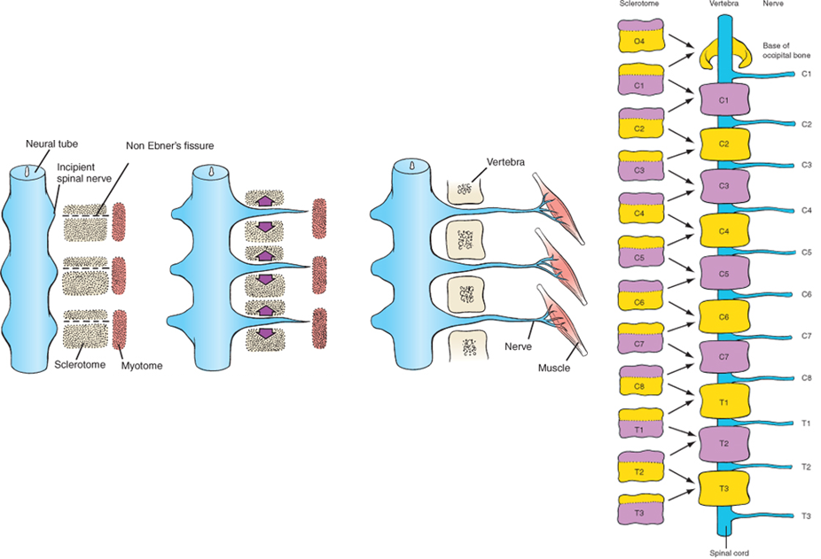
(III) Spinal cordCaudal part of neural tube

  • Neural tube has a single layer of simple columnar epithelium and central canal
  • The epithelium proliferates – forms 3 layers:
  1. Ependymal layer – produces cerebrospinal fluid (CSF) 
  2. Mantle layer – grey mater
  3. Marginal layer – white mater
development of spinal cord, neural tube

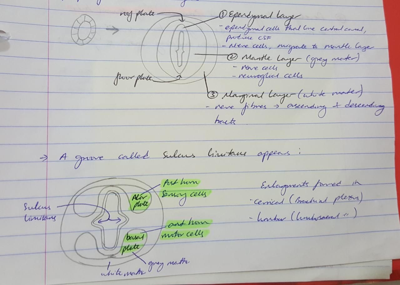
A groove called sulcus limitans divides the mantle layer into:

  1. Alar plate – posterior horn, sensory cells
  2. Basal plate – anterior horn, motor cells

Growth of spinal cord:

  1.  3rd month fetal life – till end of vertebral column
  2. Birth – till L3
  3. Adult – till L1/L2

Congenital anomalies of spinal cord:

  1. Spina bifida oculta – failure of fusion of one vertebrae dorsal part
  2. Meningocele – failure of fusion of 2-3 vertebra. meninges bulge out
  3. Meningo-myelocele – spinal cord bulges out
  4. Myelocele – Neural tube failed to close, neural plate exposed
Image result for meningomyelocele vs meningocele
Image result for myelocele
congenital anomalies of spinal cord

(IV) BrainCranial end of neural tube

  • Neural tube expands to form brain swelling
  • Lumen forms ventricles
  • 2 constrictions divide brain swelling into 3 parts

(V) Cerebellum Alar lamina of metencephalon

  • Alar lamina bend medially, forming medial and lateral bulges
  • The medial bulges meet each other over the roof plate of 4th ventricle, forming vermis 
  • Lateral bulge forms cerebeller hemispheres
  • Cerebeller cortex formed by – neuroblast cells migrating from mantle layer to marginal layer
  • Dentate nucleus – neuroblasts deeply situated in mantle layer
  • Cerebeller peduncles – axons of neurons of cerebeller nuclei grow out to reach brainstem
Image result for development of cerebellum

Development of vertebral column

  • Notochord forms
  • Sclerotome cells (paraxial mesoderm) surround notochord
  • Form mesenchymal vertebral column

Image result for caudal half of one sclerotome

  • Caudal half of one sclerotome and cranial half of the next, forms one vertebral mesenchyme, which goes through endochondral ossification  to form bone
  • Notochord regresses in regions of vertebral body
  • Notochord enlarges between the bodies to form nucleus pulposus

Image result for caudal half of one sclerotome notochord


Each vertebral body gives 3 processes:

  • 2 dorsal process – unite to form spinous process
  • 2 lateral process – in cervical region, unites with coastal process to form foramen transversarium. In thoracic region forms transverse process
  • 2 coastal process – in thoracic region elongates to form ribs

Image result for cervical and thoracic vertebrae


Development of mammary glands

  • Linear thickening of surface ectoderm (milk line) appears on ventral body
  • Extends from axilla to medial inguinal region
  • Line disappears except in pectoral region
development of mammary glands, milk line

  • 15 to 20 solid ectodermal buds arise in remaining milk line
  • Buds form solid cords
  • Which grow in underlying mesenchyme
  • Cord grows and branches in fetal life
  • Canalized before birth to form: Lactiferous sinus, Lactiferous duct, Secretory alveoli
  • Open in depressed area, which elevates after birth – to form the nipple
Image result for lactiferous sinus

At puberty:

  • Males: have ducts without alveoli
  • Females: Ovarian hormone increases branching of lactiferous duct, and increases fat between lobules. Therefore the mammary gland enlarges.

Congenital anomalies:

  1. Amastia – absence of one or two mammary glands
  2. Athelia – absence of one or two nipples
  3. Macromastia – large breasts
  4. Micromastia – small breasts
  5. Polythelia – accessory nipples
  6. Polymastia – accessory breast
  7. Gynaecomastia – male developing female breasts
  8. Klinefelter syndrome – XXY in males, therefore have enlarged breasts, wide hips and penis does not grow

Development of skin

The development of skin consists formation of:

  • Epidermis
  • Dermis
  • Subcutaneous tissue
  • Hair
  • Sweat glands
  • Sebaceous glands

(I) Epidermis:

  • Ectoderm
  • Neural crest cells – Form melanoblasts (melanin secreting cells)

(II) Dermis – paraxial mesoderm (dorsal portion) – dermatome (contributes skin, fat and connective tissue)

(III) Subcutaneous tissue – lateral plate mesoderm – somatic layer (most of the skin derived)

(IV) Hairs:

  • Solid ectodermal buds sink into underlying mesenchyme
  • Forms hair bud, which is invaginated by mesoderm
  • Forms hair papilla
  • Muscle fibers attach to hair follicles – errector pili muscles
  • Hair bulb central cells elongate and keratinize – hair shaft
  • Fetus first hair – lanugo hair
Image result for Development of hair

(V) Sweat glands:

  • Solid ectodermal buds sink into underlying mesenchyme
  • Terminal end coils and it canalizes
Image result for Development of sweat glands

(VI) Sebaceous glands:

  • Side branches of hair follicle buds
  • Central cells degenerate forming fatty secretions

Congenital anomalies:

  1. Ichthyosis – dry scaly skin
  2. Hypertrichosis – excessive hair
  3. Atrichia – congenital absence of hair
  4. Dermoid – cystic swelling along lines of fusion

Development of skeletal muscles and limbs

Skeletal muscles

Origin: Paraxial mesoderm – myotomes

Myotomes ⇒ myoblast cells ⇒ fuse ⇒ myotubes ⇒ myofilaments develop in myotubes ⇒ myotubes become myocytes (muscle cells)

somites, development of head and neck, development of skeletal muscles and limbs
  • 1st occipital myotome – extra occular muscles of the eye (NB: iris and ciliary muscle – neural ectoderm)
  • Remaining 3 occipital myotomes – muscles of tongue


somite development

The 8 cervical, 12 thoracic, 5 lumbar, 5 sacral, 8-10 coccygeal myotomes divide into:

  • Dorsal part – Epimere – forms extensor muscles of back and neck
  • Ventral part – Hypomere:
  1. Split longitudinally eg. infrahyoid muscles
  2. Split to form layers eg. intercostal muscles
  3. Ventral tips unite to form eg. rectus abdominis muscle
development of skeletal muscles and limbs

NB:

  • Myotomes migrate to limb buds to form flexor and extensor muscles
  • Pharyngeal arches give muscles in head and neck

Limbs

  • Upper limb bud forms opposite C5-T1
  • Lower limb bud forms opposite L4-S3

Each limb bud is formed of:

  • Lateral plate mesoderm – somatic layer – bones, tendons and connective tissue
  • Migrating myotomes – muscles
  • Neural crest cells – melanocytes and schwann cells
  • Which is all covered by ectoderm – skin

Development:

  • Programmed apoptosis between digital rays form fingers and toes
  • Central mesoderm – cartilagenous skeleton – ossify into bone
  • 3 segments formed in each limb bud
  • Rotation of 90 degrees around long axis:
  1. Upper limb adducted, rotated laterally, thumb lateral
  2. Lower limb adducted, rotated medially, big toe is medial

Congenital anomalies of limbs:

  1. Amelia – failure to develop limbs
  2. Phocomelia – absence of proximal limbs, direct foot or hand
  3. Polydactyl – Extra fingers or toes
  4. Syndactyl – fusion of 2 fingers or toes
  5. Claw feet – lobster deformity
  6. Congenital digit amputation
  7. Constriction band
  8. Club foot
  9. Ectodactyl – missing middle finger

Development of face and palate

Face

5 processes formed by proliferation of neural crest cells (ectoderm)

These cells migrate from mesencephalon and rhombencephalon into arches

  • 1 frontonasal process – cranial to stomodeum
  • 2 maxillary processes – lateral, from 1st pharyngeal arch
  • 2 mandibular processes – caudal, from 1st pharyngeal arch

1 frontonasal process:

  • 2 nasal placodes develop
  • Each invaginated to form:
  1. Nasal pit – forms nasal cavities – open in pharynx posteriorly
  2. Lateral nasal fold – ala of nose
  3. Medial nasal fold – fuse together to form:
  • Surface: middle part of nose, filtrum of upper lip
  • Deep: anterior upper jaw with Incisor teeth, primary palate with incisive fossa

NB: Also forms nasal septum

2 maxillary processes:

  • Grow medially
  • Fuse with medial nasal fold – form upper lip
  • Fuse with lateral nasal fold – form nasolacrimal duct
  • Fuse with mandibular process – forms cheeks
  • Forms palatine shelves

2 mandibular processes:

  • Fuse with each other medially – form lower lip and chin
  • fuse with maxillary process – forms cheeks

NB: Degree of fusion of max. and mand. processes determine width of mouth

Image result for development of face


Palate

Origin: Neural crest cells (ectoderm)

  • Formed by primary and secondary palate
  • Primary palate formed from frontonasal process (fusion of medial nasal folds)
  • Secondary palate formed from 2 palatine shelves, which arise from maxillary process
  • 2 palatine shelves fuse with each other medially, and primary palate anteriorly
  • Nasal septum descends downwards and fuses with palate in midline
  • Anterior part of palate ossifies and posterior part remains fleshy to form hard and soft palate

Anomalies of face and palate:

  1. Inclusion dermoid – cystic swellings around lines of fusion of face
  2. Macrostomia – large mouth, incomplete fusion between max. and mand. processes
  3. Microstomia – small mouth, excessive fusion between max. and mand. processes
  4. Oblique facial cleft – failure of fusion between maxillary and frontonasal process
  5. Lateral cleft upper lip – unilateral or bilateral
  6. Cleft palate – unilateral, bilateral, only soft palate or cleft uvula

Development of respiratory system

Development of larynx, trachea, lungs and esophagus

Respiratory system

Origin: Endodermal floor of pharynx behind hypobranchial eminence

1. Epithelium

  • Laryngo-tracheal groove appears in the endodermal floor of pharynx behind hypobranchial eminence
  • Edges of groove unite dividing upper foregut into esophagus and laryngo-tracheal tube
  • Laryngo- tracheal tube grows caudally:
  1. Upper end – larynx
  2. Then – trachea
  3. Lower part – 2 lung buds
  • Right lung bud divides into 3 branches (main bronchii)
  • Left  lung bud divides into 2 bronchii
  • Each bronchus – subdivisions – bronchial tree
  • Terminal/respiratory bronchioles end in alveoli, expand only after birth

2. Cartilage, muscles and connective tissue: 4th and 6th arch mesoderm

  • 4th pharyngeal arch  – thyroid cartilage, cricothyroid muscle
  • 6th pharyngeal arch – cricothyroid, arytenoid, cuneiform and corniculate cartilages, and all other laryngeal muscles

NB: Splanchnic and somatic pleura forms lungs viceral and parietal pleura

Image result for laryngotracheal groove

Maturation of lungs:

Image result for maturation of lungs embryology
Image result for maturation of lungs
embryology

Anomalies of lungs:

  • Agenesis of one or both
  • Accessory lung lobe
  • Hyaline membrane disease – alveoli cannot ventilate adequately due to absence of surfactant

Esophagus

Origin: Endoderm of foregut

  • From respiratory diverticulum to stomach swelling
  • Surrounding mesenchyme forms musculature of esophagus

Anomalies of trachea and esophagus:

  1. Tracheal-esophageal fistula – incomplete fusion of laryngo-tracheal groove
  2. Esophageal atresia – narrowing of esophagus
  3. Failure of elongation – pulls stomach up thorax – Hiatus hernia

Development of tonsils, tongue and thyroid

Development of palatine tonsils, tongue and thyroid gland

Palatine tonsils

Origin: 2nd pharyngeal arch endoderm

Endoderm proliferates into buds ⇒ Canalizes ⇒ Forms crypts ⇒ Invaded by lymphocytes

Development of palatine tonsils

Tongue

  1. Muscles of tongue – 3 occipital myotomes of paraxial mesoderm
  • (1st occipital myotome forms extraoccular muscles of eye)
  • The 3 remaining myotomes drag the hypoglossal nerve with them

2. Mucous membrane:

  • Anterior 2/3 – 1st pharyngeal arch
  • From two lingual swellings and tuberculum impar
  • Posterior 1/3 – endoderm of 3rd pharyngeal arch
  • Endoderm of 2,3 and 4 arch fuse to form Hypobronchial eminence
  • A groove divides the hypobronchial eminence into:
  1. Upper part – Posterior 1/3 tongue mucous membrane
  2. Lower part – Forms epiglottis

Anterior and Posterior fuse at sulcus terminalis ( v shape)

Development of tongue

Congenital anomalies:

  • Aglossia – absence of tongue
  • Macroglossia, microglossia
  • Bifid tongue
  • Glossoschisis – cleft tongue
  • Tongue tie (ankyloglossia) – frenulum is till tip of tongue, can’t protrude tongue

Thyroid gland

  1. Endodermal thickening around foramen cecum
  2. Forms thyroid primodium between tuberculum impar and hypobronchial eminance
  3. Thyroid primodium invaginated – bilobed diverticulum
  4. Descends infront of hyoid bone and laryngeal cartilage
  5. Connected to dorsum of tongue by thyroglossal duct
  6. Finally reaches infront of thyroid cartilage and upper trachea
  7. Thyroglossal duct obliterated
Image result for development of thyroid gland

Congenital anomalies:

  • Thyroid agenesis – absence, leads to stunted physical and mental growth
  • Lingual thyroid – Fails to descend
  • Thyroglossal cyst – thyroglossal duct unobliterated
  • Thyroglossal fistula – cyst may rupture
  • Ectopic positions – Sublingual, retrohyoid, retrosternal etc.

Development of pharyngeal (branchial) apparatus

6 mesodermal thickenings on each side of primitive pharynx:

  1. Outer ectodermal covering (Externally – 5 grooves – Pharyngeal clefts)
  2. Mesodermal core
  3. Inner endodermal lining (Internally – 4 grooves – Pharyngeal pouches)
Image result for branchial apparatus
development of branchial apparatus

Basic structure of pharyngeal arch:

  1. Mesoderm – Cartilagenous bar (forms cartilage, ligaments and bones)
  2. Mesoderm – Striated muscle (special viceral muscle of head and neck)
  3. Aortic arch artery
  4. Own nerve (motor + posttrematic sensory)
  5. Next arch’s nerve (pretrematic sensory)

NB: 5th arch disappears

Derivatives of the arches:

Ectoderm, endoderm, mesoderm derivatives and shape of each pharyngeal arch:

derivatives of pharyngeal arches, from ectoderm, endoderm, mesoderm and shape

Anomalies:

  1. Branchial cyst – along anterior border of sternocleidomastoid muscle due to failure of cervical sinus to obliterate
  2. Branchial sinus – Branchial cyst opens into skin by narrow canal
  3. Branchial fistula – Branchial cyst opens into lumen of pharynx
  4. Duplication of external auditory meatus – 1st branchial anomaly

Note: If left untreated, may become repeatedly infected and inflamed. Recurrent inflammation makes surgical resection more difficult. Excellent prognosis if lesion is completely resected. 


Development of mouth cavity and salivary glands

Mouth cavity:

  1. Floor of foregut (Endoderm)
  2. Stomodeum (Ectoderm)

In between is the Buccopharyngeal membrane which ruptures at the end of the 3rd week

development of mouth cavity


Salivary glands

1. Parotid gland: Ectodermal

  • Ectodermal groove appears behind the angle of the mouth
  • Closes to form the parotid duct
  • Branches repeatedly and canalizes to form acini
  • Duct recedes from the angle of the mouth
  • Duct opens in vestibule, opposite maxillary 2nd molar

2. Submandibular gland: Endodermal

  • Endodermal groove appears between tongue and gums
  • Closes to form submandibular duct
  • Branches repeatedly and canalizes to form acini
  • Anterior end of duct proceeds forwards , and opens close to frenulum of tongue

3. Sublingual gland: Endodermal

  • Arise as small buds from submandibular groove
  • Attain common sheath
  • Has several openings in floor of mouth

Clinicals:

  1. Cyst formation – Lymphoepithelial cysts/branchial cysts commonly found in the anterior triangle of the neck
  2. Agenesis (failure to develop) of gland
  3. Atresia (no canalization) of duct