Exogenous pigmentation
1. Accidental:
- Foreign substance – Pencil graphite
2. Iatrogenic:
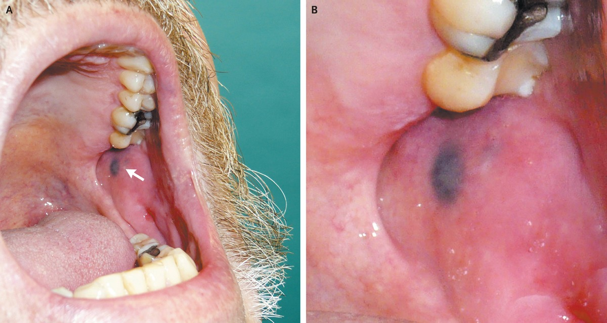
- Amalgam tattoo:
- Condensation of mercury in abraded gingiva/breakage of filling
- Histology: Dark granules along collagen bundle + multinucleated cells
- Chlorhexidine mouthwash:
- Yellow brown on surface of oral tissues and surface of teeth (cervical + interproximal)
3. Drugs and heavy metals:
- Lead & bismuth – blue black deposits along gingival margin
4. Localized:
- Hairy tongue – green brown on dorsum, overgrowth of filiform papilla by chromogenic bacteria
- More under white lesions
- Click here for picture
5. Superficial staining of oral mucosa:
- Topical medications
- Smoking
- Tobacco
- Foods/drinks
Endogenous pigmentation (melanotic lesions)
1. Developmental causes:
- Racial pigmentations
- Naevi/mole
- Peutz-Jeghers syndrome
- Polyostotic fibrous dysplasia
- Neurofibromatosis
2. Acquired causes:
- Systemic disease – Addison, HIV
- Smoking
- Hyperkeratosis and chronic inflammation/trauma
- Drugs (minocycline)
- Idiopathic oral melanotic macules
- Lentigo simplex
3. Malignant causes:
Other endogenous pigmentation
- Blood breakdown products and other disturbances of iron metabolism
1. Hemoglobin: Blue, red, purple
2.Hemosiderin/bilirubin: Brown
- Ecchymosis
- Petechia
- Hemochromatosis
- Varix/hemangioma
3. Melanin: Brown, black, grey
- Melanotic macule
- Basilar melanoma
- Naevus
- Melanoma

1 thought on “Oral pigmentations”
Comments are closed.