Viral disease
- HSV
- VZV
- Hand, foot and mouth disease
- Herpangina
Immunological disease
- Pemphigus vulgaris
- Mucous membrane pemphigoid
- Dermatitis herpetiformis
- Linear immunoglobulin disease
| Pemphigus vulgaris | Mucous membrane pemphigoid | |
| Etiology Target proteins | – Autoimmune reaction – Intercellular keratinocyte protein (desmoglein 3) destroyed by auto antibodies | – Autoimmune reaction to basement membrane proteins: – Lamina 5 – BP 180 |
| Clinical | Intraepithelial blisters | Subepithelial blisters |
| Site | – Oral mucosa – Skin – Mucosa | – Oral mucosa – Conjunctiva – Skin rarely |
| Histology | – Intraepithelial clefting – Loss of desmosomal contacts therefore free floating acantholytic Tzanck cells | – Subepithelial clefting – Chronic inflammatory infiltrate |
| Direct immunofluorescence testing | Circulating auto IgG | No circulating auto IgG |
Subtypes:
Nikolsky’s sign: Positive – stroke mucosa gently, vesicle or bulla appears
Management: Corticosteroids + Azathioprine + Cyclophosphamide
Prognosis: Good. Fatal if untreated
Management: Corticosteroids
Prognosis: Fair
Differential diagnosis:

| Minor aphthae | Major aphthae | Herpetiform aphthae | |
| Size | 0-0.5cm | >0.5cm | 0-0.5cm |
| Shape | Oval | Ragged oval, crateriform | Oval |
| Number | 1-10 | 1-5 | 10-100 |
| Location | Non keratinized mucosa | Non keratinized mucosa | Any intra oral site |

1. Systemic signs and symptoms
2. Oral infections
3. Oral neoplasms
4. Other manifestations

Age of onset: Neonate to late infancy
Infections:
Malignancy:
Nutrition:
Systemic:
Parotid: Diffuse, non suppurative enlargement
CNS: Developmental delays
AIDS dysmorphic syndrome: Variable teratogenicity
Mnemonic: SPOTS
NB: Immunocompromised patients have atypical mycobacteria infections eg. M. avium intracellulare
Mnemonic: PRIESt
Cervical TB:
Primary – Chancre (chronic ulcer) Picture
Secondary
Tertiary
NB: Can go into latency
Early
Late: Latency
Notes under white lesions
Prenatal infection:
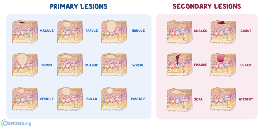
1. Macule
2. Papule
3. Nodule
4. Sessile
5. Pedunculated
6. Papillary
7. Verrucous
8. Vesicle
9. Bulla – Blister > 5mm in diameter
10. Pustule – Blister filled with purulent exudate
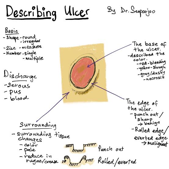
11. Ulcer
12. Erosion
13. Fissure – Narrow slit like ulceration/ groove
14. Plaque – Elevated and flat on surface
15. Petechia – Round pinpoint area of hemorrhage
16. Ecchymosis – Non elevated hemorrhage, larger than petechia
17. Telangiectasia
18. Cysts
19. Unilocular – Radiolucent lesion – single compartment
20. Multilocular – Radiolucent lesion – several compartments
Physical trauma
Therapeutic radiation
1. Acute traumatic ulcer
2. Chronic traumatic lesions
1. Neoplasm
2. Immunological disease
3. Aphthous ulcer/ Canker sore – Multifactorial
4. Infections
Nb: Chronic infectious ulcers (TB, syphilis, fungal)
Group of fibrous connective tissue (CT) lesions that occur when over exuberant repair (granulation tissue and scar) follows injury
Clinical:
Histology:
Management: Surgical excision + full mouth scaling
Etiology:
Clinical:
Etiology: Chronic trauma due to I’ll fitting denture
Site:
Clinical:
Histology:
Management:
Oral wart/ verruca vulgaris (autoinoculation from wart)
Low grade variant of OSCC
