Inferior part of pelvic outlet between thighs, separated from pelvic cavity superiorly by pelvic floor
Boundaries:
- Anterior – Pubic symphysis, mons pubis/base of penis
- Posterior – Tip of coccyx, intergluteal cleft
- Lateral – Medial thigh, inferior ischiopubic rami, sacrotuberous ligament
- Roof – Pelvic floor
- Base – skin and fascia

Blood supply: Internal and external pudendal
Nerves: Pudendal, ilioinguinal, posterior cutaneous nerve of thigh
Lymphatics:
- Glans penis/clitoris – Deep inguinal nodes
- Testis/ovaries – Lumbar
- Rest of perineum – Superficial inguinal
Perineal body:
Fibromuscular mask at junction of urogenital and anal triangle. Has skeletal muscles, smooth muscles, collagen and elastic fibers
Muscles that attach to it:
- Levator ani
- Bulbospongiosum
- Superficial and deep transverse perineal muscles
- External anal sphincter
- External urethral sphincter
Clinicals:
- Damage during childbirth – Stretching or tearing, therefore possible prolapse of pelvic viscera. Can be avoided by episiotomy (surgical cut in the muscular area between the vagina and the anus)
- Pudendal and ilioinguinal nerve block – during labour or episiotomy
Anal triangle
Contents:
- Anal aperture
- External anal sphincter muscle
- Two ischioanal fossae – spaces lateral to anus
Ischioanal/ischiorectal fossae:
Boundaries:
- Anterior – Pelvic diaphragm, perineal body
- Posterior – Sacrotuberous ligament, gluteus maximus
- Superior – Levator ani
- Inferior – Perineal skin
- Medial – Anal canal, external sphincter
- Lateral – Ischial tuberosity, obturator internus
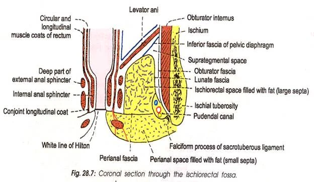
Content:
- Fat
- Internal pudendal vessels
- Pudendal nerve
- Inferior rectal vessels and nerve
Clinicals:
- Ischianal abscess – infection due to wound
- Anal fissure – anal valve tears
Urogenital triangle
Coronal section:

Superficial perineal pouch:
Boundaries:
- Anterior – continuous with Scarpa’s fascia
- Roof – Perineal membrane
- Floor – Perineal fascia
- Lateral – Ischiopubic ramus
Contents:
- Root of penis
- Superficial perineal muscles
- Ischiocavernosus and bulbospongiosum muscles
- Vestibular glands (♀)
- Superficial transverse perineal nerve
- Clitoris (♀)
- Bulb of vestibule (♀)
Deep perineal pouch:
Boundaries:
- Superior – Pelvic diaphragm
- Inferior – Perineal membrane
- Lateral – Obturator fascia
Contents:
- Deep transverse perineal muscles
- External urethral sphincter
- Membranous urethra
- Bulbourethral glands
- Internal pudendal vessels
- Artery of bulb of penis
Clinicals:
- Extravasation of urine – interruption of urethra, collection of urine in scrotum or penis
- Bartholin’s gland cyst
Pelvic diaphragm
Separates pelvic cavity (true pelvis) and perineum (genitalia and anus)
Pelvic viscera (bladder, rectum, genital organs) reside in pelvic cavity
Pierced by: Rectal hiatus, urogenital hiatus (urethra, vagina)
Functions:
- Support viscera
- Resistance to increase in intrapelvic pressure while coughing etc
- Sphincter action on urethra and rectum
- Support fetal head
Clinicals:
- Injury during childbirth – prolapse of pelvic viscera, urinary/rectal incontinence
Urinary bladder
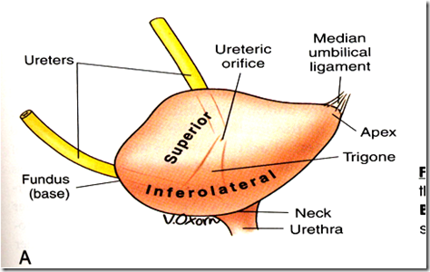
Position: Anterior in pelvic cavity, posterior to pubic symphysis, infront of uterus, cervix and vagina
2 sphincters:
1. Internal urethral sphincter:
- Males: Circular smooth fibers, autonomic control, prevent seminal regurgitation during ejaculation
- Females: No muscle
2. External urethral sphincter: Skeletal muscle under voluntary control
Relations:
- Anterior – pubic bone, median umbilical ligament
- Posterior – Rectum, vas deferens, seminal vesicle, vagina, uterus
- Superior – Peritoneum, sigmoid colon, coils of small intestine, fundus of uterus
- Inferior – Pelvic diaphragm, prostate
- Lateral – Obturator internus, levator ani muscles
Support: Median umbilical ligament, pelvic diaphragm, urogenital diaphragm, puboprostatic ligament (males) and pubovesical ligament (females)
Blood supply: Superior and inferior vesical, obturator, inferior gluteal, vaginal and uterine (for females)
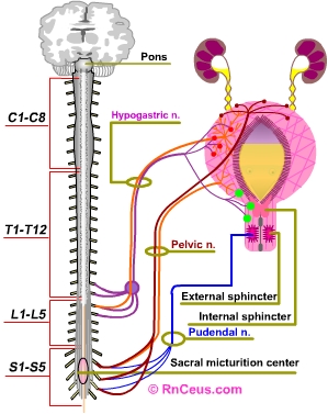
Nerves:
- Sympathetic – Hypogastric nerve (T12-L2) – relax detrusor muscle, urine retention
- Parasympathetic – Pelvic splanchnic (S2-S4) – contract detrusor muscle, stimulate micturition
- Somatic – Pudendal nerve (S2-S4) – innervate external urethral sphincter, constrict (storage), relax (micturition)
Lymphatics: Internal and external iliac
Bladder stretch reflex:

Clinicals:
- Spinal cord injury:
- Above T12 – No awareness of bladder filling, no control over external sphincter, constantly relaxed bladder
- Below T12 – Flaccid bladder, detrusor muscle paralysed, bladder fills uncontrollably
- Rupture of bladder – fracture/injury, urine escapes to extraperitoneal or intraperitoneal
- Cystocele – prolapsed bladder into anterior vagina wall
- Cystostomy – opening of bladder to drain urine
- Cystoscopy camera inserted into bladder via urethra
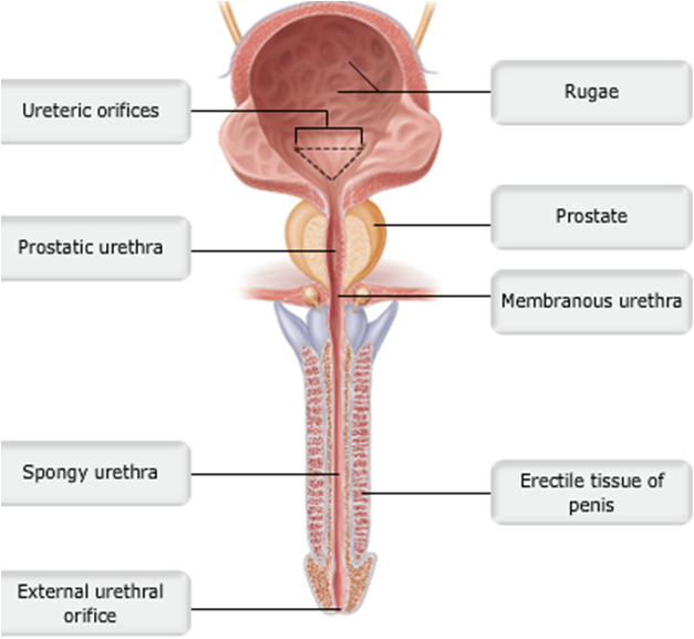
Urethra
Extent:
- Males: Internal urethral orifice (bladder) – external urethral orifice (tip of glans)
- Females: Neck of bladder – urethral orifice in vetibule
Blood supply: Inferior vesical, middle rectal, dorsal artery of penis, artery of bulb, internal pudendal, vaginal
Venous: Prostatic venous plexus, internal pudendal
Nerves: Inferior hypogastric plexus (sympathetic and parasympathetic), pudendal (somatic)
Lymphatics: Internal iliac, deep inguinal
NB: Male urethra divided into 4 parts:
- Preprostatic: Internal urethral orifice – prostrate
- Prostatic: Through prostate gland, ejaculatory duct and prostatic ducts drain into urethra here
- Membranous: Surrounded by external urethral sphincter – voluntary control
- Spongy: Through bulb and corpus spongiosum, bulbourethral glands empty here
Clinicals:
- Urinary tract infection
- Male catheterisation – insert tube through urethra into bladder when patient cannot pass urine
Rectum
Position: True pelvis, posterior end
Flexures: 2 anteroposterior and 3 lateral
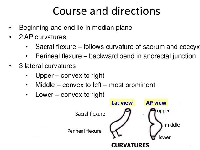
Final segment of rectum is called ampulla – relaxes to store faeces
Relations:

Blood supply:
- Superior rectal (from IMA)
- Middle rectal (from internal iliac)
- Inferior rectal (from internal pudendal)
Nerves: Hypogastric plexus
Lymphatics: Pararectal and internal iliac
Clinicals:
- Hemorrhoids – thrombosis of external rectal plexus
- Proctoscope – examine anal canal, rectum and sigmoid colon
- Rectocele
- Digital rectal examination
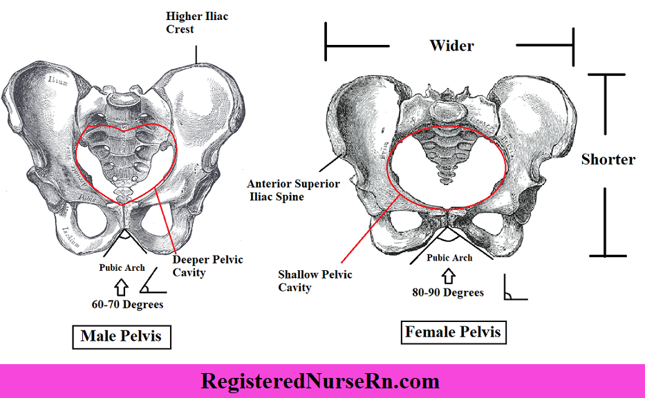
Difference between male and female pelvis
Anal canal
Position: In anal triangle between ischioanal fossae, from rectum to anus
Relations:
- Lateral: Ischioanal fossae
- Posterior: Coccyx and sacrum
- Anterior: Perineal body, urogenital diaphragm, urethra, bulb of penis, vagina
Sphincters:
- Internal – upper 2/3, involuntary
- External – Lower 1/3, voluntary
NB: Pectinate line – divides anal canal into upper (embryonic hindgut) and lower (ectoderm of proctodeum) parts
Blood supply: Superior rectal (above pectinate line), Inferior rectal (below)
Nerves: Autonomic – Inferior hypogastric plexus (above pectinate line), Somatic – pudendal nerve (below)
Lymphatics: Internal iliac (above pectinate line), superficial inguinal (below)
Clinicals:
- Anal fissure – anal valve tears
- Hemorrhoids – constipation
- Perianal abscess
- Anal fistula
- Anorectal incontinence – pudendal nerve damage
These are summarized notes from various sources, mainly TeachMeAnatomy and Wikipedia
