Bones and how to side them
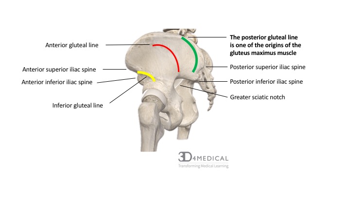
Pelvis:
To side pelvis, look at the ischial tuberosities, obturator foramen and acetabular notch


Femur:
- Posterior: Linea aspera, intertrochanteric crest
- Medial condyle larger and downwards, adductor tubercle on medial side

NB: Blood supply to head of femur – Nutrient artery, artery in ligamentum teres, medial and lateral circumflex arteries
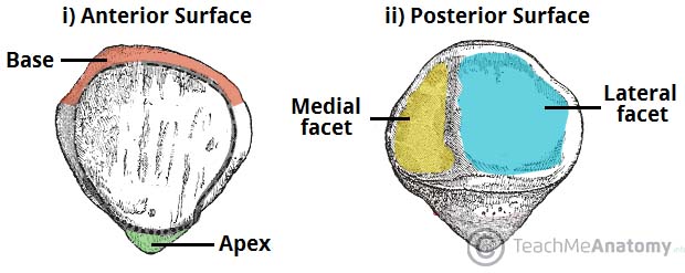
Patella:
- Ant: rough surface
- Post: smooth, lateral facet larger
Tibia:
- Anteriorly is tibial tuberosity
- Medial malleolus
- Anterior border sharpest

Fibula:
- Lateral malleolus
- Head has styloid process and articular facet on lateral head
Bones of foot:
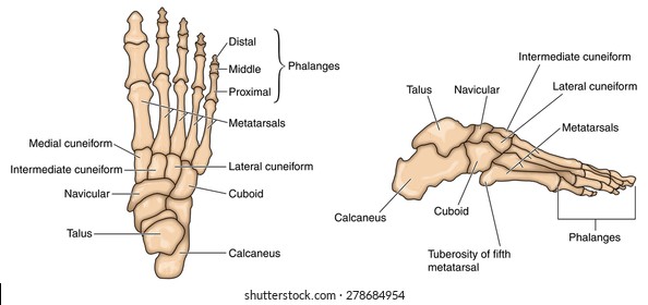
Clinicals: club foot, flat foot, hammer toe, bunion
Talus:
- On lateral side is a bulge as seen from above
- Medial surface has comma shaped articular facet
- Planter surface has deep groove
- Anterior surface has head
Cuboid:
- Proximally concave
- Superiorly broad and rough
- Inferiorly oblique groove and ridge behind the groove
- Laterally notch
- Medially oval facet and broad
Calcaneus:
- Posterior part rough and large
- Lateral surface straight, medial surface concave
Muscle attachment on bones


Arteries
(I) Anastomosis
Trochanteric anastomosis – SLIM
- Superior gluteal a.
- Lateral circumflex
- Inferior gluteal
- Medial circumflex
Cruciate anastomosis – LIMP
- Lateral circumflex
- Inferior gluteal
- Medial circumflex
- 1st perforator
Longitudinal anastomosis – perforators

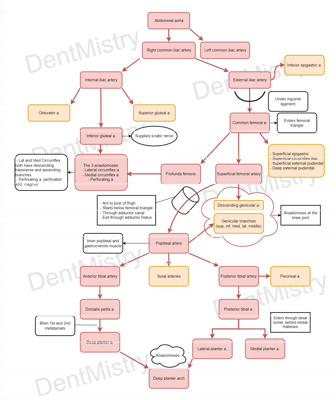
(II) Femoral artery – anterior thigh
- From external iliac artery
- Between ASIS and pubic symphysis
- Posterior to inguinal ligament
- Anterior to psoas major muscle
5 branches – Superficial circumflex iliac, superficial epigastric, superficial external pudendal, deep external pudendal and profunda femoris – lateral and medial circumflex and 1-4 perforators
- Becomes superficial femoral artery
- Descends through femoral triangle and adductor canal (in here give descending genicular artery)
- Through adductor magnus hiatus – becomes popliteal artery
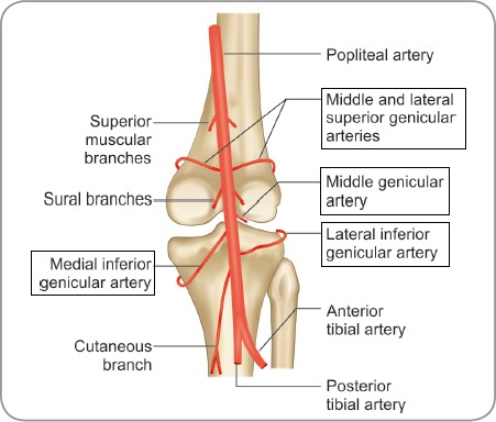
(III) Popliteal artery
- Through adductor hiatus
- Descends popliteal fossa
- Between femoral condyles
- Superficial to popliteus muscle
- At lower border of popliteus muscle branches into anterior and posterior tibial arteries
- Other genicular branches: (supply knee joint)
Clinicals – muscles compress artery
(IV) Anterior tibial artery – anterior leg and lateral leg
- Origin from lower border of popliteus muscle
- Passes anteriorly by piercing upper end of interosseous membrane
- Descends between tibialis anterior and extensor digitorum longus
- Below inferior extensor retinaculum
- Forms dorsal pedis artery – which forms deep planter artery
(V) Posterior tibial artery – posterior leg
- Gives off a peroneal artery
- Runs in posterior compartment of leg between deep and superficial muscles
- Enters tarsal tunnel behind medial malleolus
- Divides into medial and lateral planter arteries
(VI) Obturator artery – medial thigh
Veins
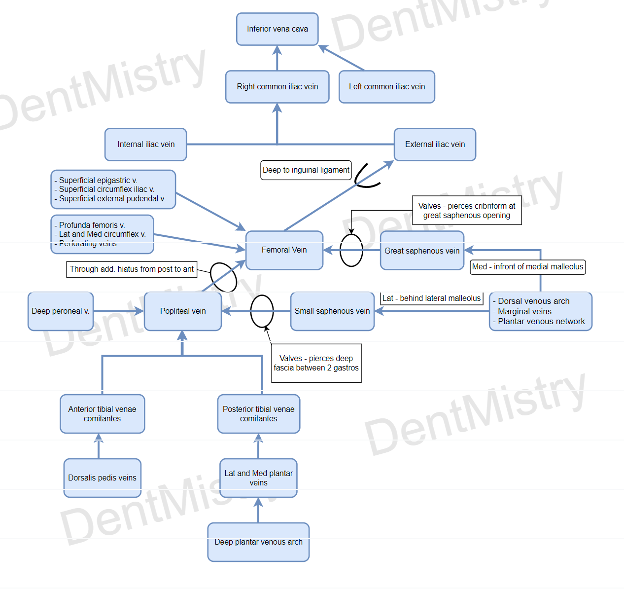
(I) Great saphenous vein
- Drains dorsal vein of big toe and dorsal venous arch of foot
- Infront of medial malleolus
- runs up medial side of leg
- Courses anteriorly
- Pierce cribriform fascia at saphenous opening – joins femoral vein in femoral triangle
(II) Small saphenous vein
- Drains dorsal vein of small toe and dorsal venous arch of foot
- lateral aspect of foot behind lateral malleolus
- Runs up posterior aspect of leg
- Pierces deep fascia between 2 gastrocnemius
- Drain into popliteal vein
Clinicals:
- Varicose veins – incompetent valves of superficial veins
- Grafts – Coronary bypass
- Venous cutdown – infuse fluid in dehydrated children at medial malleolus
Nerves
Cutaneous Innervation:
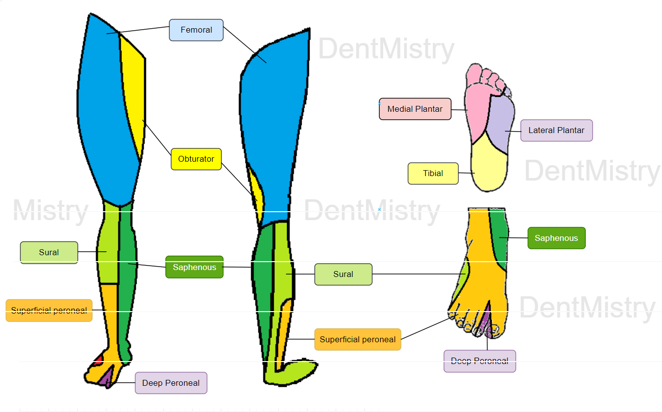
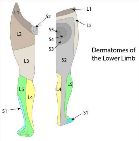
Dermatomes:
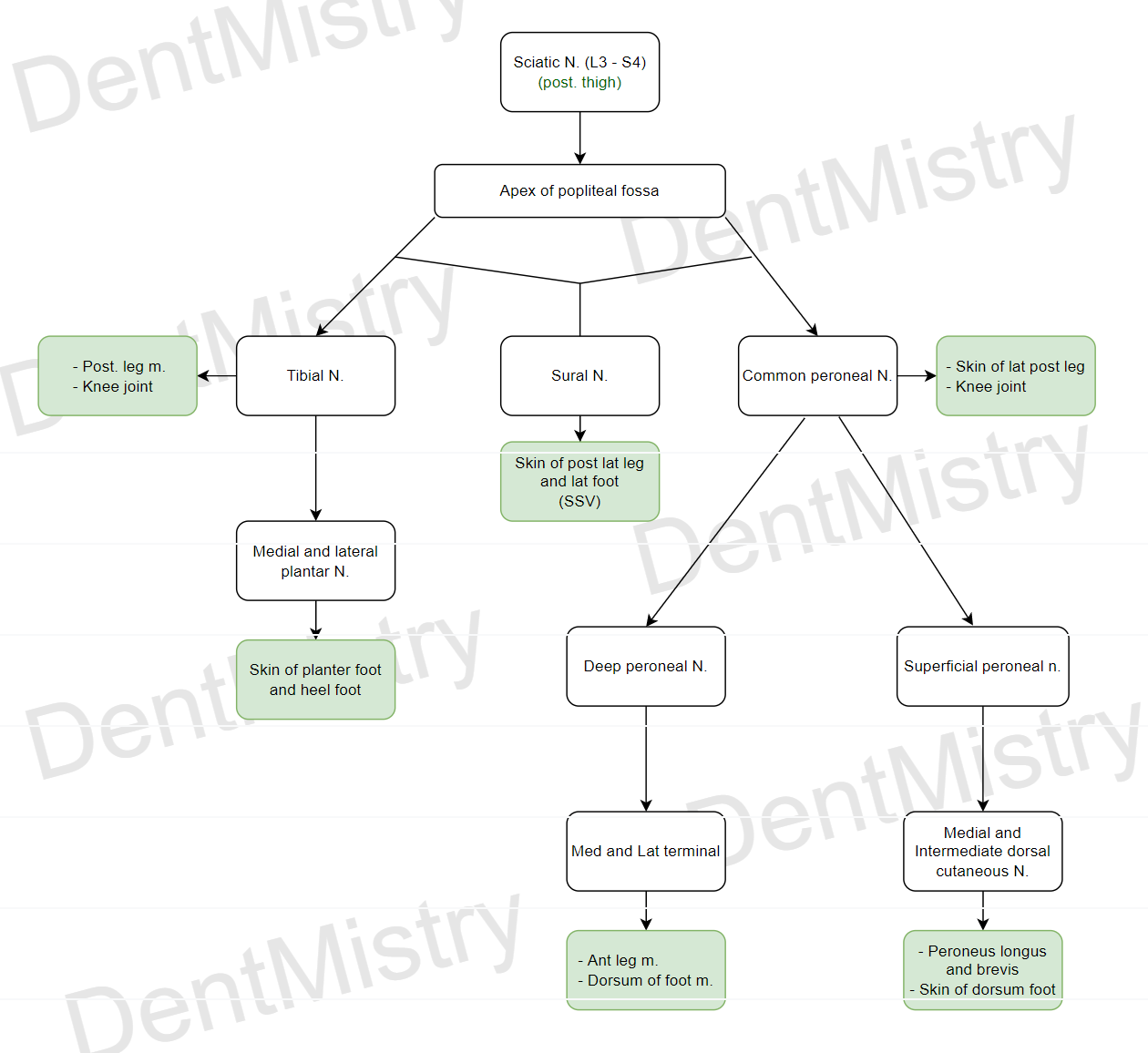
(I) Sciatic nerve: L4-S3 – posterior thigh
Course:
- Passes between imaginary line between PSIS to ischial tuberosity to gluteal tuberosity
- Through infrapiriform compartment, greater sciatic foramen
- Deep to gluteus maximus muscle
- Descends posterior thigh
- Superficial to adductor magnus
- Deep to biceps femoris
- At apex of popliteal fossa divides into tibial nerve and common peroneal nerve
Supplies – posterior thigh
Blood supply – inferior gluteal artery, perforators of profunda femoris
Clinical:
- Piriformis foramen syndrome
- Injury by wound/femur dislocation – muscles below knee paralyzed
- Foot drop – peroneal nerve
- Sciatic hernia – intestines through GSF
(II) Tibial nerve (accompanies posterior tibial artery) – posterior leg
- Contributes to sural nerve
- Runs in posterior compartment of leg between deep and superficial muscles
- Enters tarsal tunnel behind medial malleolus
- Divides into medial and lateral planter nerves
(III) Common peroneal nerve (damaged – foot drop, called policeman’s nerve)
- Descends obliquely on lateral side of popliteal fossa (along medial margin of biceps femoris)
- Winds around head of fibula
- Deep to peroneus longus – divides into deep peroneal nerve and superficial peroneal nerve
(IV) Deep peroneal nerve – anterior leg
- Runs on anterior surface of interosseous membrane with anterior tibial artery
- At ankle joint, goes through extensor retinaculum, divides into medial and lateral terminal branches
(V) Superficial peroneal nerve – lateral leg
- Lateral compartment of leg
- Superficial to peroneus brevis
- Pierces deep fascia to become cutaneous
- Divides to form medial and intermediate dorsal cutaneous nerve
(VI) Sural nerve
- Between medial and lateral gastrocnemius muscles, pierces fascia
- Descends with small saphenous vein
- Behind lateral malleolus
- Runs in lateral foot
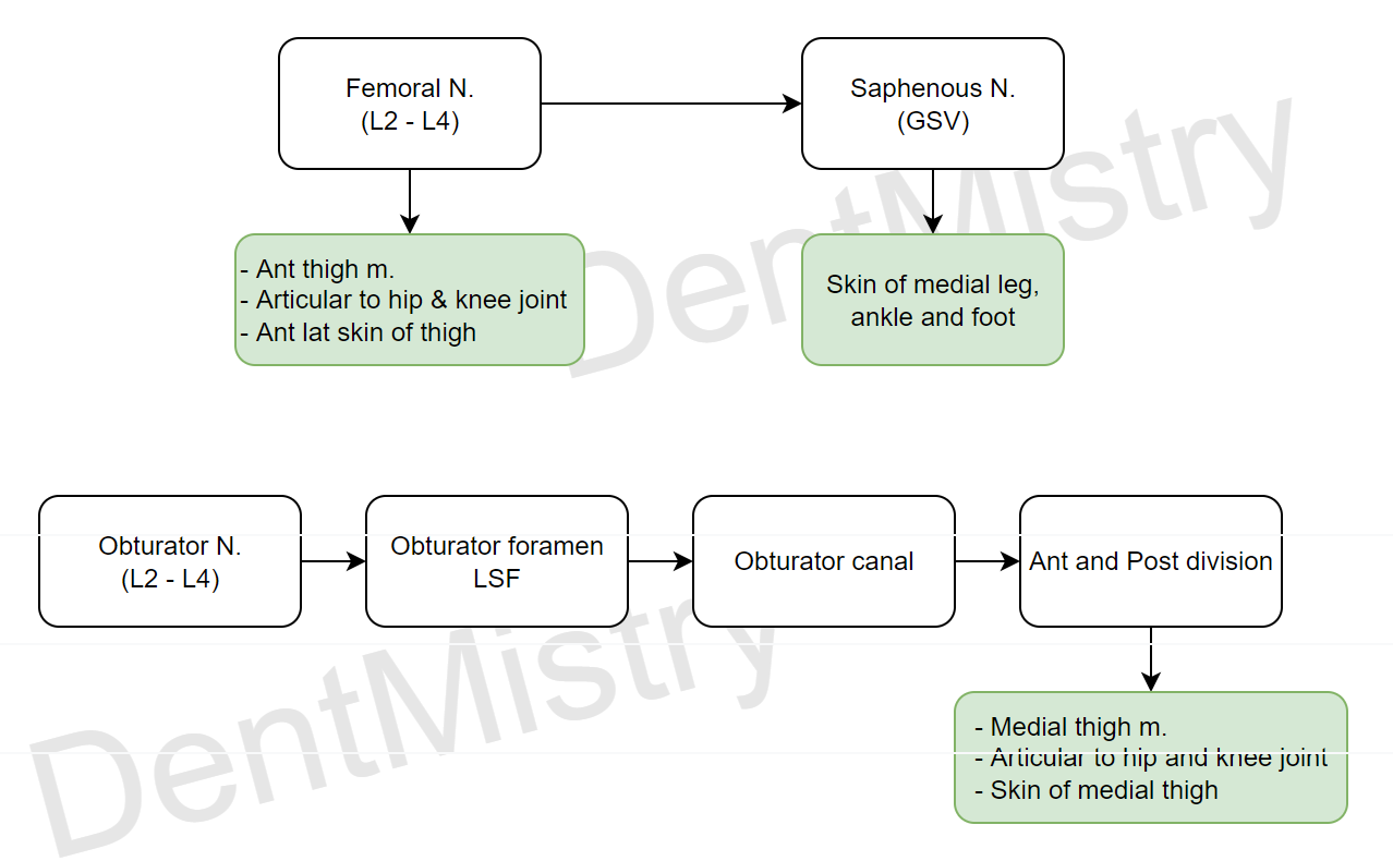
(VII) Femoral nerve: L2-L4 – anterior thigh
- From lumbar plexus
- through psoas major muscle
- Behind inguinal ligament
- Through femoral triangle outside sheath
- Splits into anterior and posterior divisions
- Terminal branch – saphenous nerve
(VIII) Saphenous nerve
- Descends through adductor canal with femoral artery and vein
- Pierces fascia, descends with great saphenous vein
- Goes through flexor retinaculum
- Runs in medial foot
(IX) Obturator nerve – medial thigh
Joints
(I) Hip joint
Classification: multiaxial ball and socket
Articular surfaces: head of femur and acetabulum notch
Stability factors:
- Static: Joint capsule, labrum, depth of acetabulum, ligaments (iliofemoral, pubofemoral and ischiofemoral)
- Dynamic: gluteus medius, gluteus minimus, iliopsoas and piriformis
Movements: flex, extend, adduct, abduct, medial rotation, lateral rotation
Blood supply: trochanteric anastomosis and nutrient artery
Nerve supply: femoral, obturator, sciatic and superior gluteal
Clinicals: hip joint dislocations, psoas bursa and femoral fractures
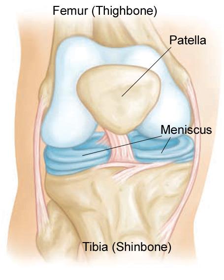
(II) Knee joint – largest joint, lined with hyaline cartilage
Classification: synovial modified hinge joint (condylar and sellar (between femur and patella))
Articular surfaces: patella and condyles of femur and tibia
Stability factors:
- Static:
- Joint capsule
- Pateller retinaculum (extensions of aponeurosis of vasti medialis and lateralis on each side of patella)
- Intercondylar eminence
- Pateller ligament (to tibial tuberosity)
- Anterior and posterior cruciate ligaments
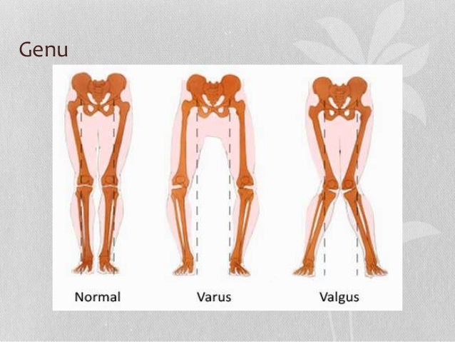
- Tibial and fibular collateral (resist valgus instability)
- Bursa – semimembranosus, suprapateller, popliteal
2. Dynamic: iliotibial tract, semimembranosus and semitendinosus muscle
Movements: flex, extend, medial and lateral rotation
Blood supply: genicular anastomosis and descending genicular artery
Nerve supply: femoral, obturator, tibial and common peroneal
Clinicals:
- Ligament tears
- Housemaids bursities – prepateller
- Clergymans bursities – infrapateller
- Unhappy triad – medial collateral, medial menisci and anterior crutiate damaged
- Pateller dislocation – laterally
- Q angle:
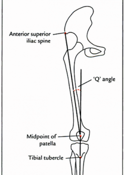

Atypical Q angles
NB: Menisci – fibrocartilage structure. Acts as shock absorber, load transmission, proprioception, produce synovial fluid, protect articular cartilage
NB: Tibia and fibula have a syndesmosis joint and therefore are immovable and are joint by connective tissue. Even radius and ulna
(III) Ankle joint
Classification: synovial hinge joint
Articular surfaces: tibia, fibula and trochlea of talus
Stability factors:
- Static: deltoid ligament, tibiofibular transverse ligament, anterior and posterior talofibular ligament, calcaneofibular ligament
- Dynamic: tendons of anterior and posterior leg muscles
Movements: dorsiflexion and planterflexion
Blood supply: malleolar branches of anterior and posterior tibial arteries and peroneal artery
Nerve supply: tibial and deep peroneal
Clinicals:
- Ankle sprains – lateral ligament weaker so inversion
- Potts fracture – eversion, breaks lateral malleolus
Others
- Walking phase:
2. Venous return in lower limbs: Valves, muscular pumps, venae comitans

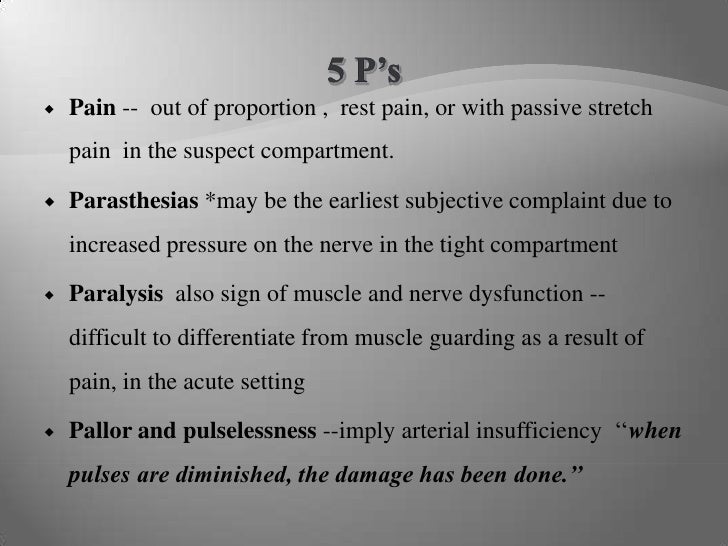
3. Compartment syndrome:
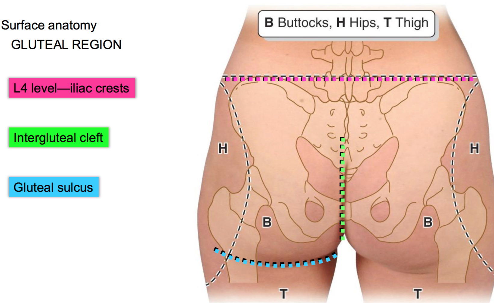
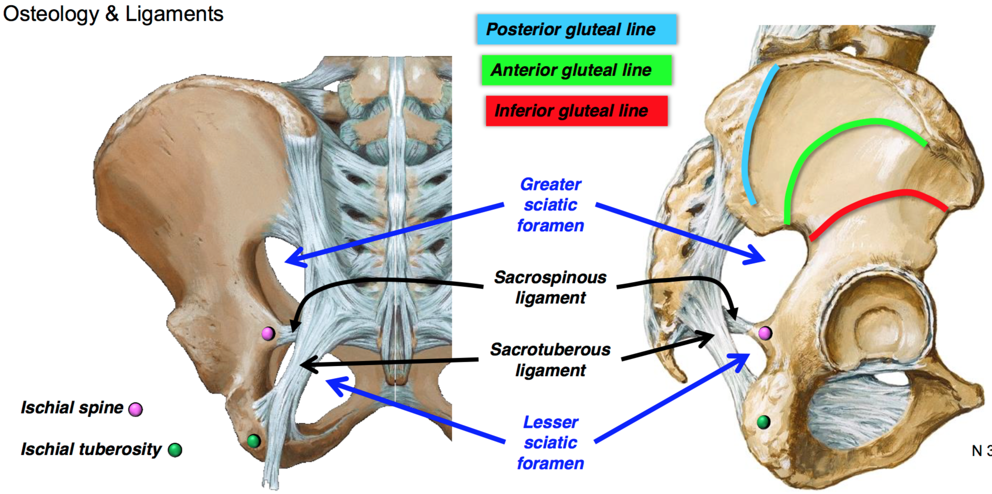
4. Extents of gluteal region:
5. Sacrospinous ligament and sacrotuberous ligament form:
(I) Greater sciatic foramen – which piriformis muscle divides into:
- Suprapiriformic – superior gluteal A,V,N
- Infrapiriformic – inferior gluteal A,V,N, pudendal nerve and sciatic nerve
(II) Lesser sciatic foramen – Pudendal nerve, nerve and tendon of obturator internus
6. Popliteous muscle – Tibia fixed, rotates femur laterally. Femur fixed, rotated tibia medially
7. Fascia lata:
Attachments:
- Superiorly – ASIS, sacrum, coccyx, iliac crest
- Inferiorly – Bones around knee
Modifications: Iliotibial tract, saphenous opening, cribriform fascia, intermuscular septa
Functions:
- Muscle attachments
- Compartmentalize
- Enclose thigh muscles – less energy used
Clinicals: Fascia lata grafts, compartment syndrome, muscular hernia if fascia cut
8. Saphenous opening – covered by cribriform fascia
Structures passing through: small saphenous vein, superficial epigastric artery, superficial external pudendal
Formed by: Cribriform fascia (roof) and falciform margin
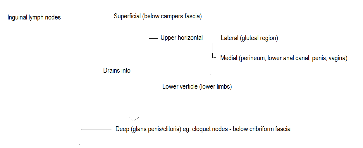
9. Inguinal lymph nodes
10. Iliotibial tract: from iliac crest to lateral patella and lateral condyles
Function: muscle attachment, stabilize lateral knee, maintain hyperextended knee position
Clinical: iliotibial band syndrome – lateral knee pain
11. Angle of declination


12. Leg compartments


13. Triceps surae – gastrocnemius and soleus muscle. Blood supply- sinusoidal
Tendon achilles : blood supply – water shed
14. Pes anserinus – conjoined tendons of sartorius, gracilis and semimembranosus insert on upper medial tibia
15. Tendons from medial to lateral going through the extensor retinaculum:
- Tom – tibialis anterior
- Has – Extensor hallucis longus
- A – Anterior tibial artery
- Very – vein
- Nice – deep peroneal nerve
- Dog – extensor digitorum longus
- Pet – peroneus tertius
16. Tarsal tunnel contents from medial to lateral
- Tom – tibialis posterior
- Dug – flexor digitorum longus
- A – posterior tibial artery
- Very – vein
- Narrow – tibial nerve
- Hole – flexor hallucis longus
Clinical: tarsal tunnel syndrome – compressed tibial nerve
17. Popliteal fossa:
Boundaries:
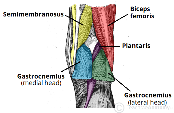
- Roof – skin and fascia
- Floor – knee joint capsule, popliteus muscle
Contents:
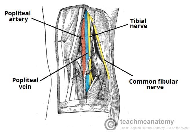
Clinical: Baker’s cyst (semimembranous bursa)
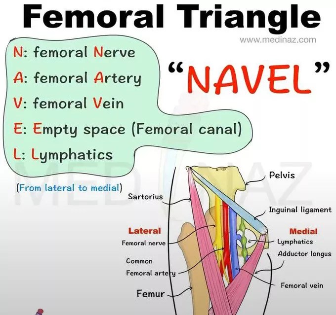
18. Femoral triangle:
Boundaries:
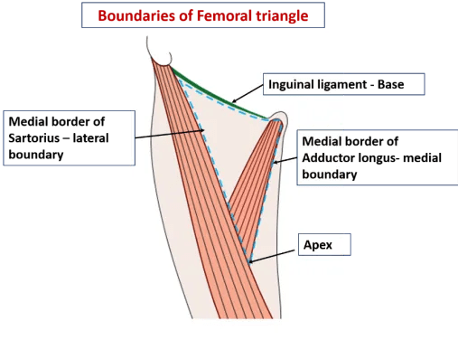
- Roof – skin and fascia
- Floor – iliopsoas, adductor longus and pectineus
Contents:
- Femoral ring and sheath
- Femoral artery, genitofemoral nerve and femoral vein (in the sheath)
- Femoral nerve
- Inguinal lymph nodes
Clinicals: femoral hernia, enlarged lymph nodes
19. Femoral ring:

Formation:
- Anteriorly – Fascia transversalis
- Posteriorly – Fascia iliacus
Clinical: femoral hernia
20. Adductor canal
Boundaries:

Contents:
- Femoral artery and vein
- Descending genicular artery
- Nerve to vastus medialis
- Saphenous nerve
Clinical: Adductor canal compression syndrome (hypertrophy of muscles)
These are summarized notes from various sources, mainly TeachMeAnatomy and Wikipedia
