Category Archives: Oral Pathology

Gardner syndrome

Etiology:

AD – mutation in adenomatous polyposis coli (APC) gene – located on chromosome 5q21

Clinical:

Triad:

  • Potential malignant colorectal polyposis
  • Skeletal abnormality – multiple osteoma
  • Multiple impacted teeth, supernumerary teeth and odontomas
Gardner syndrome clinical presentation

Diagnosis:

  • Blood test – APC gene mutation
  • Osteomas and impacted teeth found on radiograph – do lower GIT endoscopy for colon polyps

Management:

  • Alleviate symptoms
  • Surgical excision of osteomas

Fanconi syndrome

Etiology:

AR – Familial aplastic anemia

Clinical:

  • Kidney tubule dysfunction – no reabsorption. Glucose, bicarbonate, phosphorous, uric acid, potassium increased excretion in urine
  • Strabismus
  • Low set ears
  • Hypopigmentation
  • Abnormal or absent thumbs
  • Dental anomalies
  • Increased predisposition to squamous cell carcinoma

Management:

  • Bone marrow replacement
Dental association with fanconi syndrome

Ehlers-Danlos syndrome

Etiology:

Mutations lead to:

  • Defect in collagen and procollagen structures
  • Defective collagen synthetic enzymes

Clinical:

Hyper elasticity of skin and joints, fragile blood vessels

1. Systemic:

  • Bleeding tendencies
  • Poor wound healing
  • Rupture of vital organs

2. Skin:

  • Excess skin on palm and toes
  • Subcutaneous fatty cysts
  • Molluscoid pseudotumors

3. Oral:

  • Enamel hypoplasia
  • Severe PDL disease
  • Dislocation of TMJ

4. Eyes:

  • Ectopic lentis (displaced lens)

NB: Patients who cannot metabolize copper suffer from this disease

Down Syndrome

Etiology:

Trisomy 21:

  • Non-disjunction (94%)
  • Translocation
  • Mosaicism

Risk increases with mothers age

Clinical:

1. CVS:

  • Ventricular septal defect
  • AV communication
  • Persistent ductus arteriosus
  • Mitral valve prolapse
  • Teratology of Fallot:
    • Pulmonary valve stenosis
    • Ventricular septal defect
    • Overriding aorta
    • Right ventricular hypertrophy
Teratology of fallot

2. CNS:

  • Delayed motor function
  • Early aging
  • Mental retardation
  • Dementia

3. Eyes:

  • Epicanthic folds
  • Convergent strabismus
  • Nystagmus (involuntary eye movement)
  • Refractive errors
  • Keratoconus (conical cornea)
  • Cataracts
  • Ocular hypertelorism
Down syndrome facial features

4. Oral:

  • High palatal vault
  • Insufficient soft palate
  • Macroglossia
  • Mouth breathing
  • Delayed eruption
  • Hypodontia
  • Microdontia
  • Malocclusion

5. Immunity:

  • Impaired
  • Neutrophil defects
  • Thyroid dysfunction
  • Increased risk of ALL (Acute lymphocytic leukemia)

6. Musculoskeletal:

  • Underdeveloped maxilla and sphenoid
  • Prognathic mandible
  • Rib and pelvic abnormalities
  • Subluxation of patella
  • Simian palmar crease
Simian palmer crease

Diagnosis:

In utero:

  • Ultrasound
  • Amniocentesis
  • Chronic villus sampling
  • Blood test
  • Nuchal translucency test

New born:

  • Blood test – chromosomal karyotype

Management:

Regular reviews for; CVS, audiologic, ophthalmologic, dental

Crouzon syndrome/ Craniofacial dysostosis

Crouzon syndrome, craniofacial dysostosis

Etiology:

  • AD – missense mutation in FGFR2 gene
  • Premature fusion of cranial sutures – increased intracranial pressure – therefore prevents skull from growing normally and affects shape of head and face

Clinical:

Cranial deformities:

  • Frontal bossing
  • Premature fusion of coronal suture
  • Brachycephaly
  • ‘Hammered silver’ appearance of skull

Facial malformations:

  • Maxilla hypoplasia
  • Mandible prognathism

Dental deformities:

  • High arched palate
  • Peg laterals
  • Partial anodontia

Eye deformities:

  • Exophthalmos
  • Divergent strabismus
  • Blindness
  • Neuritis
  • Hypertelorism

Others:

  • Spina bifida occulta

Management:

  • Surgical
  • Orthodontic treatment
Hammered silver appearance of skull meaning

Clefts of lips and palate

Etiology:

Many contributing risk factors: Genes & environment

  • Genetic predisposition
  • Syndromes associated with cleft li and palate
  • Nutrition: iron, zinc, folic acid, B12 deficiency
  • Drugs: phenytoin, valproic aid
  • Alcohol
  • Maternal smoking
  • Stress and disturbance during development

Cleft lips: Failure of medial & lateral nasal process to be penetrated by epithelial cells during 6th week in utero.

Cleft palate: Lack of lateral palatal segment fusion – ingrowth of mesoderm – and epithelial breakdown during 8th week in utero

Results in – Mastication, speech, deglutition defect

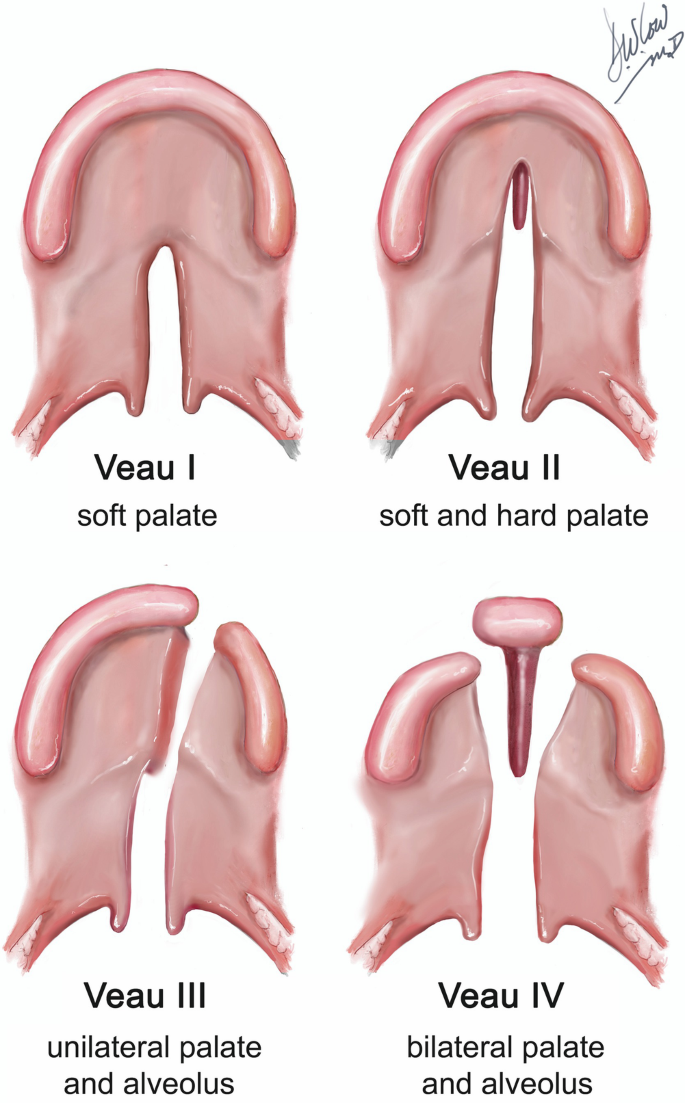
Veau classification:

Cleft lips:

Class IUnilateral notch of vermillion border, not extending to lip
Class IIUnilateral cleft of lip, not extending to floor of nose
Class IIIUnilateral cleft of lip, extending to floor of nose
Class IvBilateral cleft of lip
Cleft lips classification table
Veau classification of cleft lip

Cleft palate:

Class IUnilateral cleft limited to soft palate
Class IIUnilateral cleft of hard and soft palate – not extending past incisive foramen (2ry palate)
Class IIIUnilateral cleft extending from uvula to alveolar process
Class IvBilateral cleft of soft and hard palate
Cleft palate classification table
Veau classification of palate

Diagnosis:

  • Ultrasonography for baby in utero

Management:

  • Lip surgery
  • Palate repair – before speech development (1 and half years age)
  • Speech therapy
  • Alveolar bone grafting
  • Orthodontic treatment
  • Orthognathic surgery (jaw exercise)

NB:

  • Age 1-3 months – Lip taping and naso-alveolar molding
  • Age 3 months – Repair of cleft lip
  • Age 9-12 month – Repair of cleft palate
  • Age 1-7 years – Orthodontic treatment
  • Age 7-8 years – Alveolar bone graft
  • 18 years old or skeletal maturity – Midface advancement, continued orthodontic treatment

Rule of 10’s:

  • Weight ≥ 10 pounds
  • Hemoglobin ≥ 10 mg/dl
  • Age ≥ 10 weeks

Associated syndromes:


Van der Woude syndrome

  • Lower lip pits in patient with cleft lip and palate
  • AD
  • Mutations in interferon regulatory factor 6 gene (IRF6)
  • Associated features:
    • Hypodontia
    • Molar incisor hypomineralization
    • Ankyloglossia (tongue tie)
    • Syndactyly (webbed fingers)

Picture

Cherubism

  • Genetic and fibro-osseous disease
  • Developmental jaw condition
  • 100% penetrance in male, 50-75% in female (M:F = 2:1)
  • Mean age 7 (1 – 12 years age)

Etiology:

  • AD – mutation of SH3BP2 on chromosome 4
  • Hyperactive macrophages and osteoclast – lead to bone loss and inflammation

Clinical:

  • Teeth displaced and loose
  • Painless bilateral expansion of mandible and maxilla (starts at 2-4 years)
  • Therefore ‘cherubic’ exposure of sclera

Diagnosis:

X-ray:

  • Multilocular expansile radiolucency – soap bubble appearance (ie. multilocular cysts)
  • Opacification of sinuses

Histology:

  • Vascular fibrous tissue with multinucleated giant cells
  • Bone formation progresses as lesion matures

Serology:

  • Normal calcium and phosphorus levels (to differentiate from Brown’s tumor)
  • Increased alkaline phosphatase (due to increased bone turnover)

Management:

  • Involution after puberty
  • Surgery if persistent bone deformity

Differential diagnosis:

Picture

Cleidocrania dysplasia/ dysostosis

Etiology:

  • AD – microdeletion defect of transcription factor (CBFA-1) in chromosome 6
  • Failure in intramembranous & endochondral ossification

Clinical:

Clavicle:

  • Aplasia/ hypoplasia of 1 or both clavicles

Skull:

  • Enlarged transverse diameter of cranium
  • Short sagittal diameter of cranial base
  • Delayed closure of fontanelles & sutures
  • Wormian bones in suture lines
Cleidocranial dysplasia - open fontanelle

Teeth:

  • Multiple supernumeraries
  • Retrusion of maxilla
  • Delayed eruption of permanent teeth

Diagnosis:

Radiograph:

Clavicle:
  • Aplastic/ hypoplastic
Skull:
  • Patent fontanelles & wormian bones
  • Broad cranial sutures
  • Underdeveloped nasal sinuses
Teeth:
  • Multiple, unerupted supernumeraries

Histology:

  • Teeth lack 2ry cementum (cellular cementum at apical two thirds)

Management:

  • Protect with headgear in early life (open fontanelles)
  • Surgical exposure of unerupted teeth + orthodontic therapy
  • Orthognathic surgery
  • Prosthesis

Burning mouth syndrome/ Glossodynia

Burning mouth syndrome

Usually in menopausal women

Clinical:

  • Pain and burning sensation in mouth
  • Dysgeusia (altered taste)
  • Xerostomia

Diagnosis & treatment:

  1. Candidiasis – Brush/ incisional biopsy – Antifungals
  2. Xerostomia – Drug/ anxiety history – Oral lubricants
  3. Nutritional deficiency – Levels of vitamin B, iron, zinc – Dietary supplements
  4. Abnormal tongue habit – History – Topical corticosteroids
  5. Depression – Psychiatry – Tricyclic antidepressants
  6. Pernicious anemia – Specialist
  7. DM – Specialist
  8. Hormonal imbalance – Specialist