

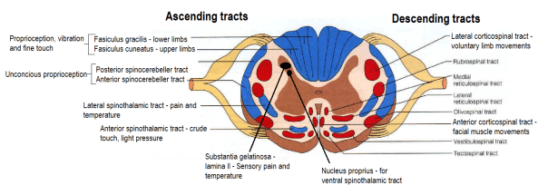
Ascending pathways
- 1st, 2nd and 3rd order
- Lesions before decussation – ipsilateral paralysis
- Lesions after decussation – contralateral paralysis
(I) Pain and temperature – Lateral spinothalamic
- Receptors:
- Free nerve endings – pain
- Krause end bulb
- Ruffini’s corpsules
- 1st order neuron
- Dorsal root ganglion
- Spinal cord dorsal horn
- Synapse with 2nd order neuron in substantia gelatinosa
- Decussate in anterior white commisure
- Ascend as lateral spinothalamic tract
- And then as spinal lemniscus in medulla
- To VPLN of thalamus
- Synapse with 3rd order neuron
- Posterior limb internal capsule and corona radiata
- Posterior central gyrus
(II) Crude touch and pressure – Ventral spinothalamic
- Receptors:
- Paccinian corpuscle – pressure
- Merkel’s disc – mechanoreceptor
- 1st order neuron
- Dorsal root ganglion
- Spinal cord dorsal horn
- Synapse with 2nd order neuron in nucleus proprius
- Axons rise a few segments
- Decussate in anterior white commisure
- Ascend as anterior spinothalamic tract
- And then as spinal lemniscus in medulla
- To VPLN of thalamus
- Synapse with 3rd order neuron
- Posterior limb internal capsule and corona radiata
- Posterior central gyrus
(III) Concious proprioception, vibration and fine touch – Fasciculus gracilis and cuneatus
- Receptors:
- Paccinian corpuscle
- Muscle spindle
- Golgi tendon organ
- Meissner’s corpuscle – light touch
- 1st order neuron
- Dorsal root ganglion
- Ascend in ipsilateral fascicle
- Synapse with 2nd order neuron in medulla in nucleus gracilis/cuneatus
- Pyramidal decussation
- Ascend in medial lemniscus
- To VPLN of thalamus
- Synapse with 3rd order neuron
- Posterior limb internal capsule and corona radiata
- Posterior central gyrus
(IV) Unconcious proprioception/ reflex – From muscles to cerebellum
Ventral spinocerebeller
- Receptors:
- Muscle spindle
- Golgi tendon
- 1st order neuron
- Dorsal root ganglion
- Spinal cord dorsal horn
- Synapse with 2nd order neuron in dorsal horn
- Decussate in anterior white commisure
- Ascend as ventral spinocerebeller tract
- Enter cerebellum in superior cerebeller peduncle
- Decussate in cerebeller white mater
- Terminate in vermis
NB: No 3rd order neuron, 2 decussations
Dorsal spinocerebeller
- Receptors:
- Muscle spindle
- Golgi tendon
- 1st order neuron
- Dorsal root ganglion
- Spinal cord dorsal horn
- Synapse with 2nd order neuron in Clarke’s nucleus/ nucleus dorsalis
- Ascend ipsilateral in dorsal spinocerebeller tract
- Enter cerebellum in inferior cerebeller peduncle
- Terminate in vermis
NB: No 3rd order neuron, no decussations, ipsilateral tract
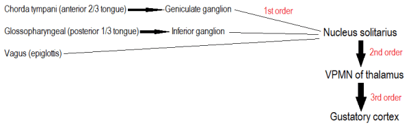
(V) Gustatory pathway – Taste
Descending pathways
- Upper motor and lower motor
Voluntary and fine movements

These are summarized notes from various sources, mainly TeachMeAnatomy and Wikipedia
