Source: Angiogenetic cells (mesodermal)
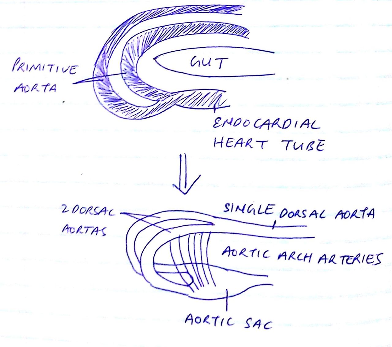
Continuation of the 2 endocardial heart tubes to form right and left primitive aorta
- Which curve dorsally and continue as 2 dorsal aortas (dorsal to gut). Cranially are separate, caudally fuse to form a single dorsal aorta
- And ventral to pharynx fuse – Aortic sac
The 2 dorsal aortas connect with the aortic sac via aortic arch arteries
These are 6 pairs of arteries that run in the 6 pharyngeal arches
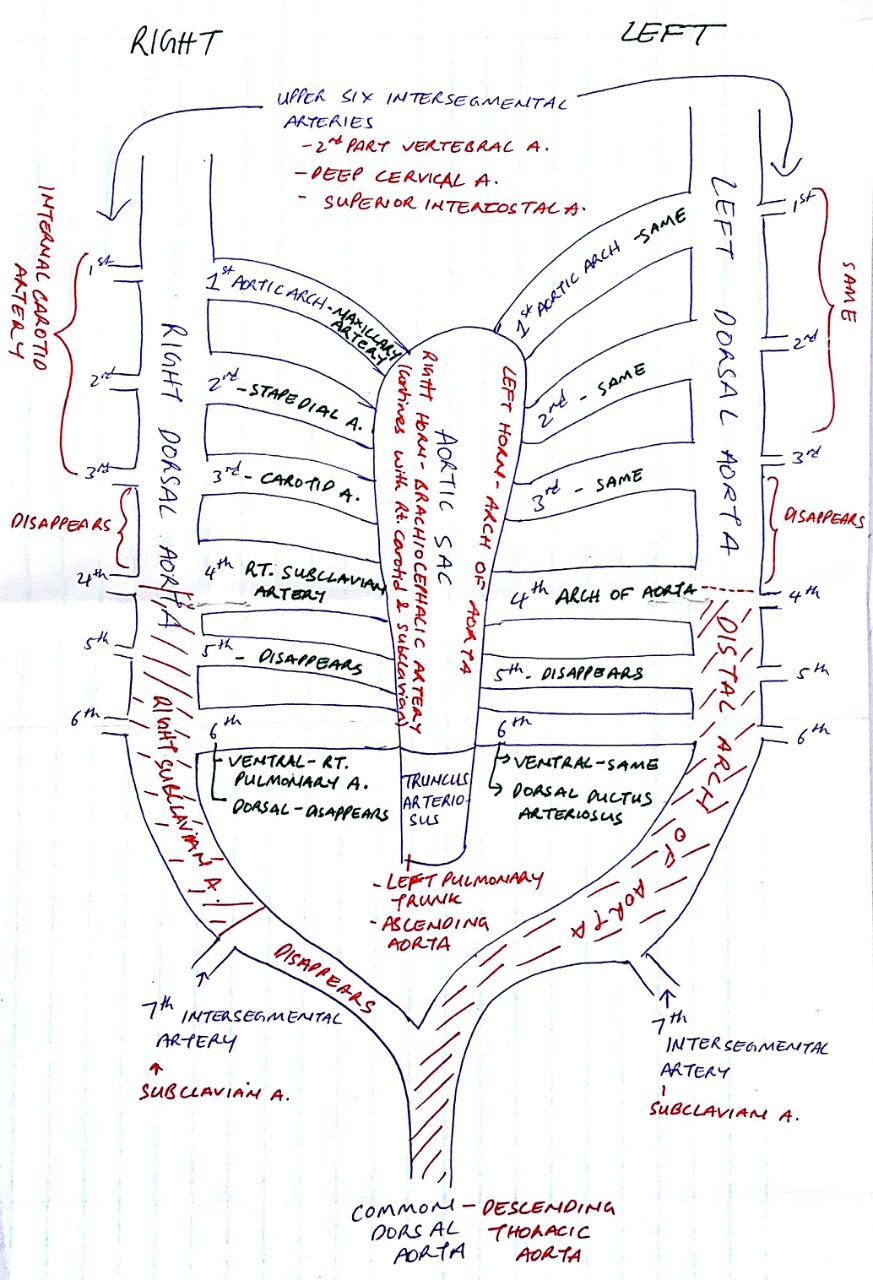

Development of arch of aorta:
- Proximal – Aortic sac stem
- Middle – Aortic sac left horn
- Distal – Left 4th aortic arch, lower left dorsal aorta
Recurrent laryngeal nerve:
- Right and left vagi descend lateral to pharynx
- Give recurrent laryngeal nerve
- On right side, 5th and 6th arch disappear (6th arch dorsal branch), therefore hooks around 4th aortic arch ie. subclavian artery
- On left side, hooks around dorsal branch of 6th aortic arch ie. ductus arteriosus
Anomalies of aortic arch:
- Patent ductus arteriosus – failure to close, hypertrophy of left side of heart
- Congenital narrowing of aorta – Preductal type (above ductus arteriosus which closes) and Postductal type (below ductus arteriosus which remains open)
- Right sided aortic arch – Distal part of left dorsal aorta degenerates, distal part of right dorsal aorta persists
- Double aortic arch – both persist, leads to dysphagia (difficulty in swallowing) and dyspnea (difficulty in breathing)
- Abnormal right subclavian artery – 4th aortic arch degenerates, therefore arises from right 7th intersegmental artery and descending aorta
Common dorsal aorta branches:
1. Ventral:
- Coelic artery – foregut
- Superior mesenteric artery – midgut
- Inferior mesenteric artery – hindgut
2. Lateral (paired):
- Middle suprarenal artery
- Renal artery
- Gonadal artery
3. Posterolateral (paired):
- Posterior intercostal arteries
- Subcostal arteries
- Lumbar arteries
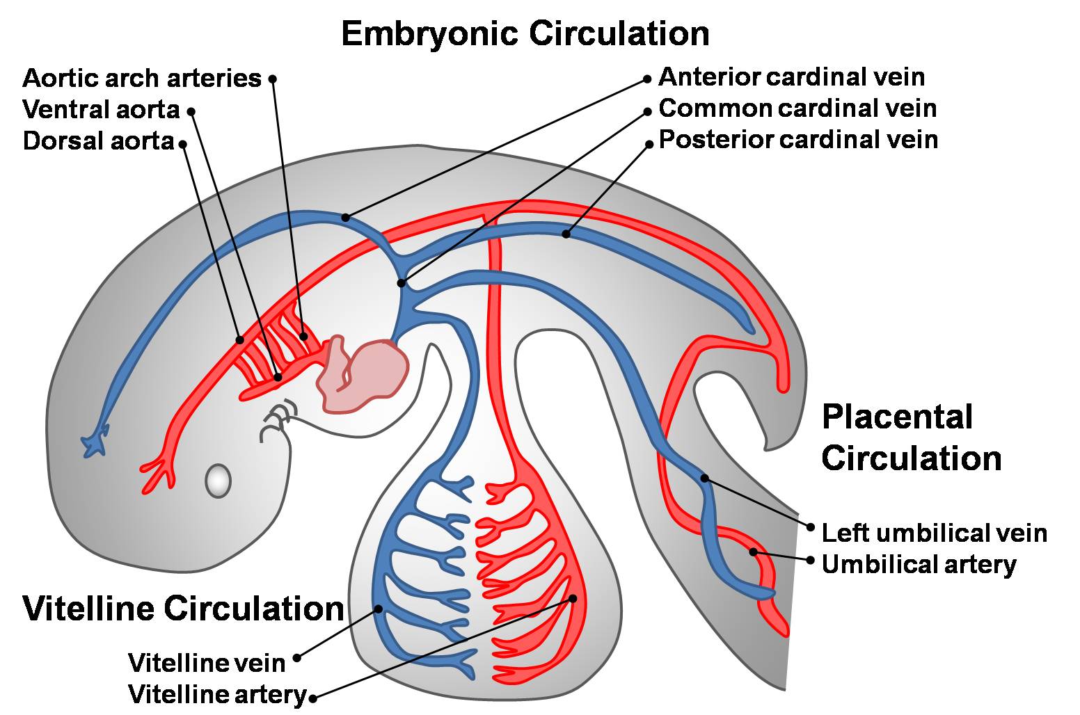
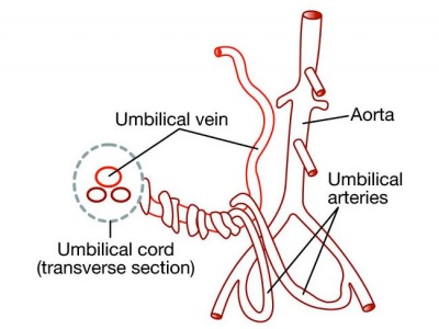
Umbilical artery:
- Arises from dorsal aorta
- Connected to 5th lumbar intersegmental artery
- Looses connection to dorsal aorta
- 5th lumbar intersegmental artery gives a branch ie. External iliac artery
- Continues as internal iliac artery
Development of arteries in lower limb:
Continuation of external iliac artery ⇒ Femoral artery ⇒ descends infront of thigh ⇒ curves to join sciatic artery backwards to form Poplitial artery
Continuation of Internal iliac artery ⇒ Called sciatic artery ⇒ descends in back of lower limb ⇒ to sole of foot ⇒ degenerates to form Inferior gluteal artery, peroneal artery ETC
EIA and IIA anastomose to form anterior and posterior tibial arteries

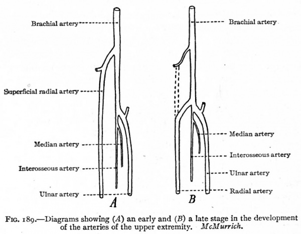
Development of arteries in upper limb:
7th cervical intersegmental artery ⇒ Subclavian a. ⇒ Axillary a. ⇒ Brachial a. ⇒ Ulnar and Radial a. ⇒ give superficial and deep palmer arches
NB: Brachial a. also gives another branch called anterior interosseous artery. Anterior interosseous artery is replaced by median artery which is replaced by ulnar artery