Amnion
Made of amnioblasts and somatopleuric layer of extraembryonic mesoderm
The connecting stalk is only made of extraembryonic mesoderm
The amnion cavity obliterates extraembryonic coelom

Functions of amniotic fluid (made from amnioblasts and fetal urine):
- Cushions the baby
- Develops the suckling reflex
- Space for urine discharge
- Maintains constant temperature
- Antiseptic – cleanses vagina when the water breaks
- Allows movement of embryo – muscle development
- Bag of waters – dilates cervix gently
Abnormalities:
- Oligohydramnios – less amniotic fluid, adhesion of embryo to itsself and the amnion
- Polyhydramnios – more amniotic fluid, premature rupture
Umbilical Cord
Tubular sheath of amnion from placenta to umbilicus (naval)
Development of the cord:
1. Primitive umbilical ring – line of reflection between amnion and ectoderm
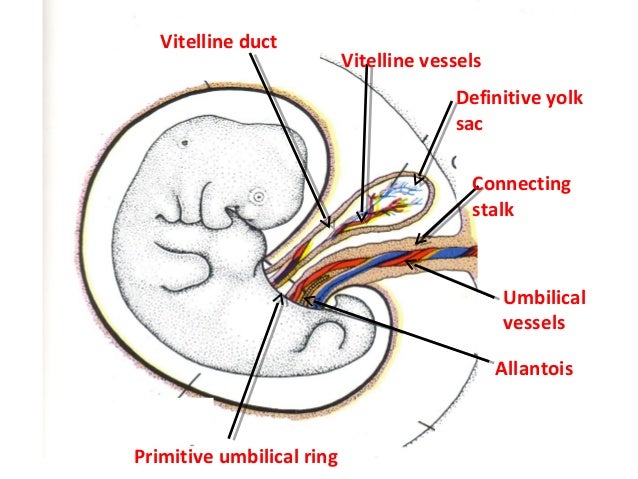
2. Primitive umbilical cord
- Body stalk
- Yolk sac
- Part of allantois (later in the chapter)
3. Definitive umbilical cord
- Wharton’s jelly (mucoid substance from extraembryonic mesoderm)
- 2 umbilical arteries
- 1 umbilical vein
- Physiological hernia between 6th and 10th week
Abnormalities:
- Short cord
- Long cord – can wrap around fetus neck
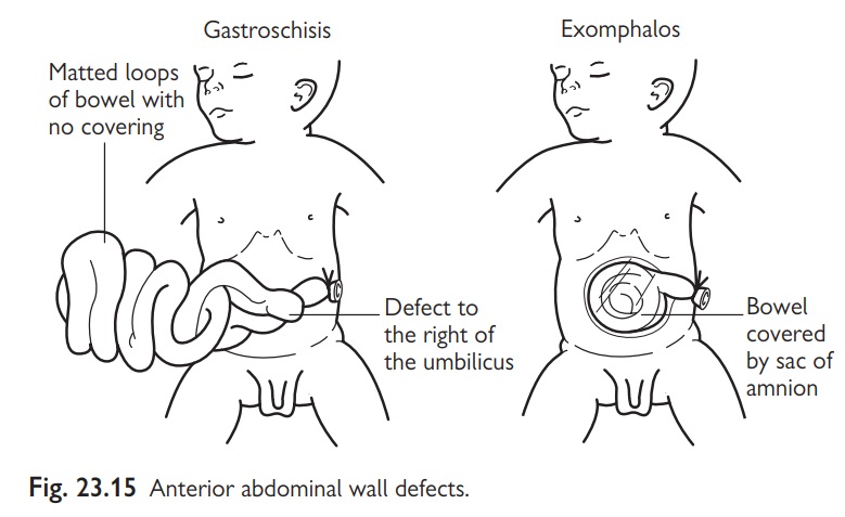
- Exomphalos – failure to reduce physiological umbilical hernia
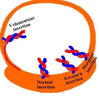
- Attachment to placenta can be eccentric, marginal or velamentous (surrounding fetal membranes)
- One umbilical artery instead of two
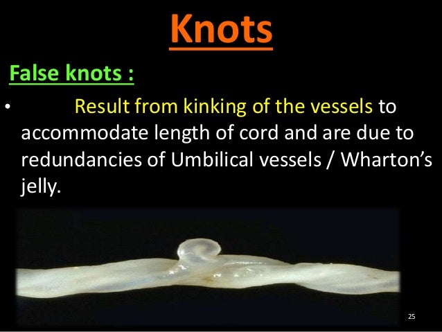
- False knots
- True knots – dangerous, obstruct blood flow
- Two to three umbilical cords
Yolk Sac
1. Primary yolk sac – Heusers membrane
2. Secondary yolk sac – Extraembryonic coelom
3. Folding of embryonic disc – primitive gut and definitive yolk sac
The embryonic disc folds in a cranial-caudal and lateral direction, because the central area of the disc grows more then the periphery. This results in the incorporation of the yolk sac roof into the embryo forming the primitive gut which is divided into foregut, midgut and hindgut.
The part of the yolk sac which was not incorporated is known as definitive yolk sac and is connected to the primitive gut by vitellointestinal duct at the midgut.
Vitelline vessels – network of vessels develop in splanchnopleuric mesoderm covering the secondary yolk sac
Functions of yolk sac:
- Roof forms primitive gut
- Caudal end forms allantois
- Gives primordial germ cells which migrate to developing gonads
- Some vitelline vessels form embryonic vessels
Allantois
Allantoic vessels form the umbilical artery and vein
It’s a tubular invagination of the secondary yolk sac
It has 2 parts:
1. Intraembryonic part: forms urachus which connects urinary bladder to umbilicus. After birth, the urachus is obliterated to form median umbilical ligament
2. Extraembryonic part: obliterated

