All posts by DentMistry

Paraaxial, intermediate, lateral plate mesoderm

The intraembryonic mesoderm differentiates into:

  1. Paraaxial mesoderm
  2. Intermediate mesoderm
  3. Lateral plate mesoderm
Image result for intraembryonic mesoderm

Paraaxial mesoderm

Transverse grooves appear dividing it into somites, from cranial to caudal end.

42 to 44 somites form;

  • 4 occipital
  • 8 cervical
  • 12 thoracic
  • 5 lumbar
  • 5 sacral
  • 8 – 10 coccygeal
Somites

The somites then differentiate in a cranio-caudal direction into:

Image result for differentiation of somites
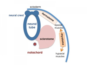
  1. Sclerotome – forms connective tissue, cartilage cells and bone cells of axial skeleton 
  2. Myotomes – differentiate into myoblasts, which then forms skeletal muscles of the body
  3. Dermatome – forms dermis of skin

Intermediate mesoderm

  • Cervical – segmented
  • Thoracic – partially segmented
  • Lumbar – unsegmented. forms nephrogenic cord (source of urinary system)

Origin to:

  • Cortex of suprarenal gland
  • Nephrons
  • Gonads

Lateral plate mesoderm

Image result for lateral plate mesoderm

Due to formation of intraembryonic coelom, it is divided into:

1. Somatic layer:

  • Adherent to ectoderm
  • Forms muscles and connective tissue of body wall

2. Splanchnic layer:

  • Adherent to endoderm
  • Forms serous membranes: Pleura, Pericardium, Peritoneum
  • Smooth muscles
  • Connective tissue of GIT, respiratory tracts
  • Cardiovascular system

The intraembryonic coelom forms:

  • Pericardial cavity
  • Peritoneal cavity
  • Pleural cavity

Other derivatives of the mesoderm:

  • All skeletal, smooth and cardiac muscles
  • Bone marrow and blood cells
  • Spleen
  • Lymph nodes
  • Dura matter

3rd week of pregnancy

In the 3rd week:

  1. Intraembryonic mesoderm forms
  2. Notochord forms
  3. Chorionic villi form

Intraembryonic mesoderm

This results in the formation of trilaminar germ disc from bilaminar germ disc. This is known as gastrulation

Gastrulation results in the formation of :

  1. Ectoderm (faces amniotic cavity)
  2. Mesoderm
  3. Endoderm (faces yolk sac)

In summary:

  1. Ectodermal cells in caudal end of bilaminar germ disc
  2. Form primitive streak, growing cranially
  3. Ectodermal cells insinuate themselves between ectoderm and endoderm
  4. Forming the intraembryonic mesoderm

Formation of notochord

  1. Primitive node and primitive pit form – thickening of ectoderm at the cranial end of primitive streak, where a pit forms in the middle
  2. Cells proliferate and a solid rod of cells grows in cranial direction between ectoderm and endoderm – known as notochordal process
  3. Notochordal process canalizes to form notochordal canal
  4. Notochordal canal fuses with underlying endoderm to form notochordal endodermal plate
  5. Which breaks down to form neurenteric canal – temporary connection between amniotic cavity and yolk sac
  6. Remaining part of notochordal plate folds along longitudinal axis. forms a solid rod of cells, known as definitive notochord
  7. Acts as a temporary axial skeleton

Formation of notochord

In summary

  1. Primitive node and primitive pit
  2. Notochordal process
  3. Notochordal canal
  4. Notochordal endodermal plate
  5. Neurenteric canal
  6. Notochordal plate folds
  7. Definitive notochord

NB: The midline region, prochordal plate and cloacal membrane do not contain mesodermal cells and remain bilaminar.

The prochordal plate will later become the buccopharyngeal membrane (oral membrane)

Formation of chorionic villi

  1. Primary villi: Syncytiotrophoblast – cytotrophoblast
  2. Secondary villi: Syncytiotrophoblast – cytotrophoblast – extraembryonic mesoderm
  3. Tertiary villi: Syncytiotrophoblast – cytotrophoblast – extraembryonic mesoderm – afferent and efferent capillaries

Primary chorionic villi

There are two types of villi:

  • Anchoring villi – penetrates into decidua basalis and anchor chorion vesicle to wall of uterus
  • Absorbing villi – branches in intervillus space

Image result for primary chorionic villi

Anchoring villi:

Anchoring villi

2nd week of pregnancy

Also known as the week of two’s:

1. Two embryoblast layers form:

  • Endoderm (Hypoblast)
  • Ectoderm (Epiblast)

Therefore forming a bilaminar germ disc

2. Two trophoblast layers form:

  • Cytotrophoblast
  • Syncytiotrophoblast – has lacunae Image result for lacunae in syncytiotrophoblast

3. Two cavities form:

  • Amniotic cavity – the cytotrophoblast cells surrounding the amniotic cavity become amnioblast cells
  • Primary yolk sac – endodermal cells grow down, line cytotrophoblast forming Heuser’s membrane (exocoelomic membrane)

Heuser’s membrane/exocoelomic membrane – Short lived combination of hypoblast cells and extracellular matrix

Image result for heuser's membrane
syncytiotrophoblast
cytotrophoblast

4. Two extraembryonic mesodermal layers form:

In the cytotrophoblast, extraembryonic mesoderm forms, in which lacunae appear and fuse to form extraembryonic coelom. Therefore dividing the extraembryonic mesoderm into 2 layers.

  • Somatopleuric mesoderm – lines cytotrophoblast and amniotic cavity
  • Splanchnopleuric mesoderm – lines primary yolk sac
Image result for splanchnopleuric mesoderm

NB: 

  • Secondary yolk sac also forms, when a large part of primary yolk sac separates and disappears leaving the smaller part. It is completely lined with endoderm.
Secondary Yolk Sac
  • Primary chorionic villi forms – cytotrophoblast projects into syncytiotrophoblast (more in the next chapter)

Syncytiotrophoblast – Placental barrier between maternal and fetal blood

Menstruation cycle and 1st week

menstruation cycle

Ovarian Cycle: Follicular phase – Ovulation – Luteal phase

Uterine Cycle: Menstruation – Proliferation – Secretory

Image result for menstruation cycle hormones

1. Follicle Stimulation Hormone (FSH)

  • Produced by anterior pituitary gland
  • Stimulates primary follicles to develop

2. Estrogen

  • Produced by primary follicles
  • Endometrium to proliferate
  • Stop production of FSH so that other primary follicle don’t develop
  • Stimulate production of LH (Luteinizing Hormone)

3. Luteinizing Hormone (LH)

  • Produced by anterior pituitary gland
  • Stimulates ovulation
  • Rest of Graffian follicle forms corpus luteum – produces progesterone

4. Progesterone

  • Maintain thick endometrium (secretory phase of endometrium)

NB:

  • If fertilized, corpus luteum will form corpus luteum of pregnancy – produces progesterone and is maintained by gonadotropic hormones produced by the embryo until placenta takes over to produce progesterone
  • If not fertilized, corpus luteum will form corpus albicans which will then degenerate
  • Reduction in production of progesterone leads to menstruation
  • Reduction in production of estrogen leads to FSH production
  • Proteolytic enzymes prevent coagulation of blood during menstruation
  1. Germinal period – 2 weeks; formation of 3 germ layers
  2. Embryonic period – 3rd week to 3rd month; differentiation of the 3 germ layers into organs and systems
  3. Fetal period – growth of various organs and systems

1st week after fertilization

Zygote →Cleavage division → Morula → Blastocyst → Hatching (degeneration of zona pellucida) → Late blastocyst

Image result for 1st week after fertilization

Inner cell mass forms embryoblast

Outer cell mass forms trophoblast; which has 2 poles:

  • Embryonic pole – faces site of implantation
  • Abembryonic pole – opposite pole

Image result for embryonic pole and abembryonic pole
blastocyst

Process of Implantation:

  1. Trophoblast erodes endometrium
  2. Penetration defect in endometrium
  3. Blastocyst embedded in endometrium of uterus
  4. Penetration defect closed by fibrin clot

Clinicals:

  1. Ectopic Pregnancy – implantation can occur in the abdomen, ovarian and fallopian tube
  2. Blighted ovum – dead embryoblast
  3. Downs syndrome – Trisomy of chromosome 21
  4. Turners syndrome – monosomy of x chromosome
  5. Triple x syndrome

Fertilization in mammals

  • Fusion of sperm nucleus with egg nucleus to form a diploid zygote cell
  • Occurs in upper parts of fallopian tube ie. Ampulla

Image result for ampulla of fallopian tube
parts of fallopian tube

Process of Fertilization

1. Ejaculation in vagina

2. Capacitation – removal of glycoproteins to make sperm active and identify ovum

3. 1st contact – Acrosome reaction

  • Contact of head of sperm with zona granulosa cells
  • Acrosome reaction – Acrosome burst by exocytosis
  • Releases hydrolytic enzymes

4. Break down of first barrier – Corona radiata

  • Enzymes digest the sticky substances that holds the follicle cells together
  • Cause cells to break away from zona pellucida

5. Break down of 2nd barrier – Zona pellucida

  • Another acrosomal enzyme then digests the zona pellucida to create path for entry of the sperm cell

6. Contact with third barrier – Oocyte membrane

Formation of fertilization membrane:

  • Head of 1st sperm makes contact with oocyte membrane
  • Oocyte membrane become permeable to calcium ions
  • Calcium ions rush into oocyte by diffusion
  • Calcium ions stimulate rapid exocytosis of cortical granules, which release chemical substances
  • Cortical reaction – release of cortical granules
  • A new membrane forms, called fertilization membrane
  • Fertilization membrane prevents polyspermy (entry of many sperm cells)

Q) How does fertilization membrane prevent polyspermy?

Acrosomal enzymes released by other sperms cannot digest the changed zona pellucida/fertilization membrane

7. Release of sperm nucleus into the secondary oocyte

8.  This stimulates 2ry oocyte to complete 2nd meiotic division and form a true ovum

9. Fusion of male and female pronucleus (enlarged nucleus) to form a zygote

Image result for corona radiata oocyte
Process of fertilization

Definition of terms

Embryology: study of developmental process from a single cell to a baby in months

Embryogenesis: formation and development of an embryo

Ontogeny: Process of development of an organism

Conceptus: Product of conception

Cleavage: series of mitotic cell divisions of the zygote resulting in blastomeres

Morula: a mass of 12 to 32 blastomeres

After a while, the morula develops a cavity and we call it a;

Blastocyst: structure formed after a cavity forms within the morula.

Blastocele: fluid that enters the cavity of blastocyst

Blastocele

Embryoblast: inner cell mass of blastocyst

Trophoblast: outer cell mass of blastocyst

Endometrium: inner epithelial lining of the uterus

Implantation: attachment and embedding of the blastocyst to the endometrium

Decidua: endometrium during pregnancy

Bilaminar disc: 2 layered embryo, occurs during second the 2nd week

Gastrula: trilaminar disc embryo (3 layered embryo)

ie. ectoderm, mesoderm, endoderm

Gastrulation: process of formation of the trilaminar disc, begins on 15th day

Neurulation: process of formation of neural tube, the primordium of the central nervous system

Primordium: beginning or first discernible indication of an organ or structure

Organogenesis: formation of body organs

Morphogenesis: process of shape development

Fetus: unborn offspring

Abortion: expulsion of an embryo or fetus before its viability (capable of living outside the uterus)

Teratology: study of birth defects

Teratogen: an agent that causes congenital (born with) defects

Anomaly: structural abnormality

Malformation: abnormal developmental process