Canine localization
- Parallax in horizontal plane: Two IOPA or USO + IOPA
- Parallax in vertical plane: OPG (↑8°) + USO (↓65-70°) to horizontal plane
- SLOB: Same Lingual Opposite Buccal
- If in line with arch, will not move
- Can do CBCT
Third molars
X-rays used:
- IOPA – difficult due to gagging
- OPG
- Oblique lateral view
- Lower/upper oblique occlusal view – buccolingual position
- CBCT
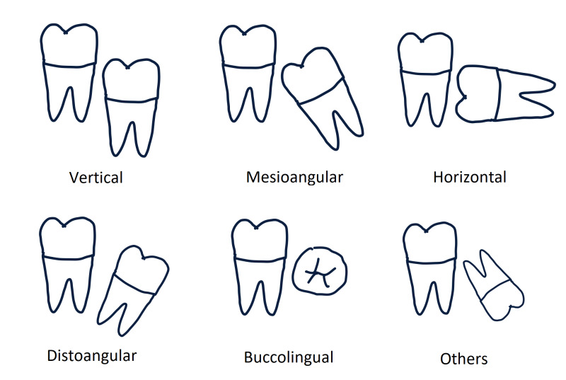
1. Angulation
2. Crown
- Size
- Shape
- Dental caries
- Resorption
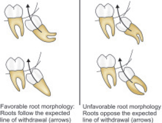
3. Roots
- Number of roots
- Shape
- Stage of development
- Curvature – favorable/unfavorable
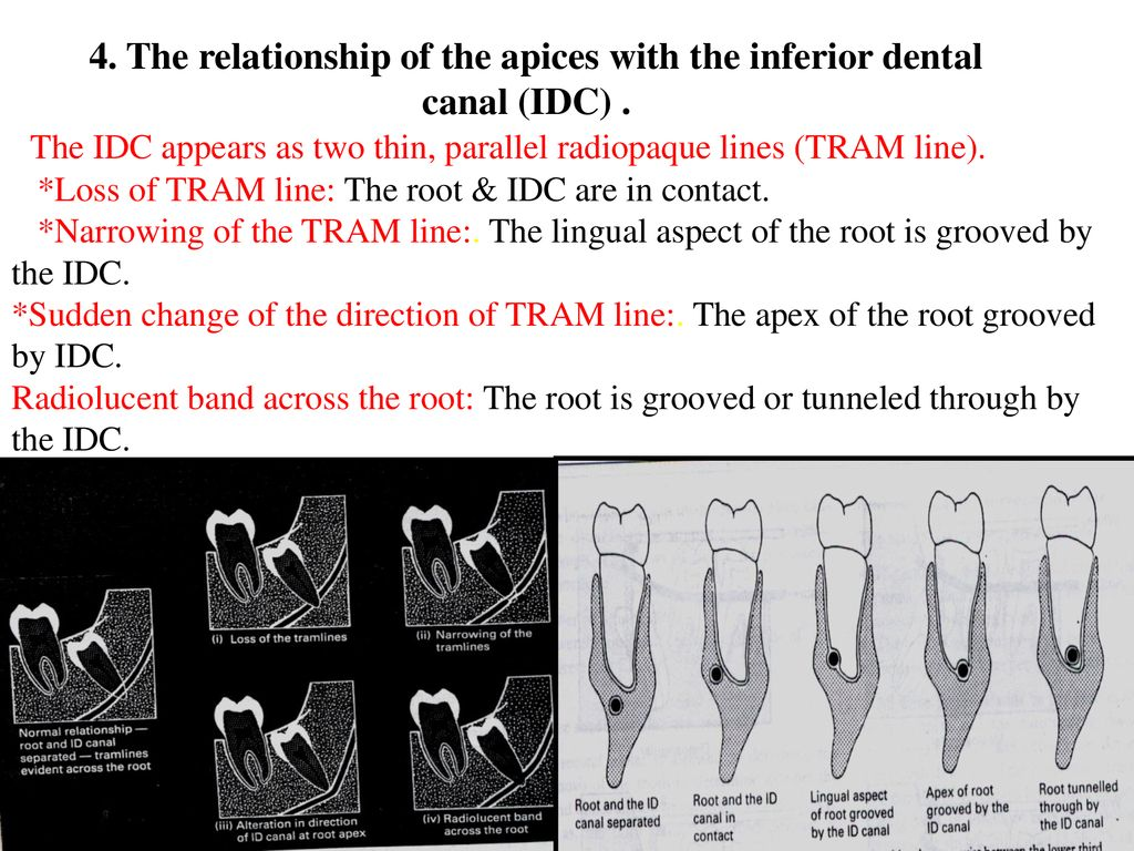
4. Relation to ID canal
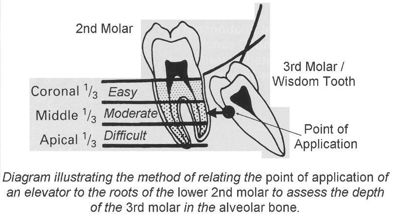
5. Depth of tooth in alveolar bone
a. CBCT measurement tools
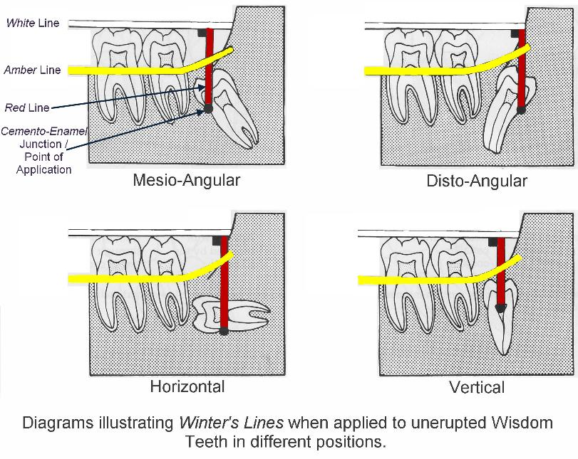
b. Winter’s lines method
- 1st line (white): Occlusal surface of 1st and 2nd molar
- 2nd line (amber):
- Crest of interdental bone between 1st and 2nd molar
- Extending distally along internal oblique ridge
- Indicates amount of investing bone surrounding the tooth
- 3rd line (red):
- Perpendicular line dropped from white line to point of application
- Measured from amber line to point of application
- If red line > 5mm – difficult extraction
c. Using roots of 2nd molar
6. Buccal or lingual obliquity
- Determine line of tooth in horizontal plane
- Buccal obliquity – Crown inclined towards cheek
- Lingual obliquity – Crown inclined towards tongue
- Use:
- Lower oblique occlusal
- Lower 90° occlusal view
7. Others
a. Surrounding bone
- Position of ascending ramus to determine access of tooth and the overlying bone
- Density of bone
- Evidence of pericoronal infection
b. Lower 2nd molar
- Crown – Condition of restoration, caries, resorption
- Root – Number, shape, periodontal status, condition of apices
