Category Archives: Embryology

2nd week of pregnancy

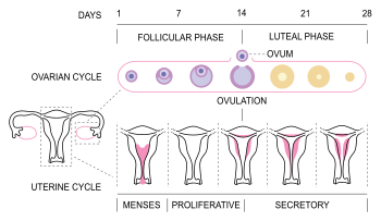
Also known as the week of two’s:

1. Two embryoblast layers form:

  • Endoderm (Hypoblast)
  • Ectoderm (Epiblast)

Therefore forming a bilaminar germ disc

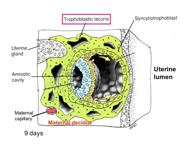
2. Two trophoblast layers form:

  • Cytotrophoblast
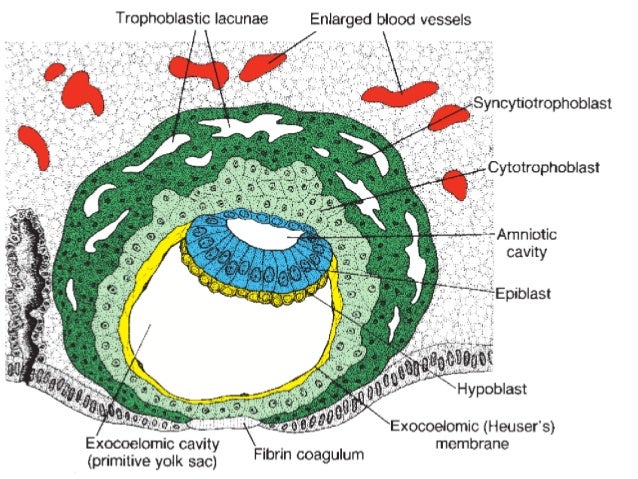
  • Syncytiotrophoblast – has lacunae Image result for lacunae in syncytiotrophoblast

3. Two cavities form:

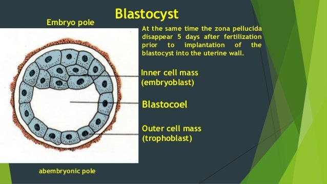
  • Amniotic cavity – the cytotrophoblast cells surrounding the amniotic cavity become amnioblast cells
  • Primary yolk sac – endodermal cells grow down, line cytotrophoblast forming Heuser’s membrane (exocoelomic membrane)

Heuser’s membrane/exocoelomic membrane – Short lived combination of hypoblast cells and extracellular matrix

Image result for heuser's membrane
syncytiotrophoblast
cytotrophoblast

4. Two extraembryonic mesodermal layers form:

In the cytotrophoblast, extraembryonic mesoderm forms, in which lacunae appear and fuse to form extraembryonic coelom. Therefore dividing the extraembryonic mesoderm into 2 layers.

  • Somatopleuric mesoderm – lines cytotrophoblast and amniotic cavity
  • Splanchnopleuric mesoderm – lines primary yolk sac
Image result for splanchnopleuric mesoderm

NB: 

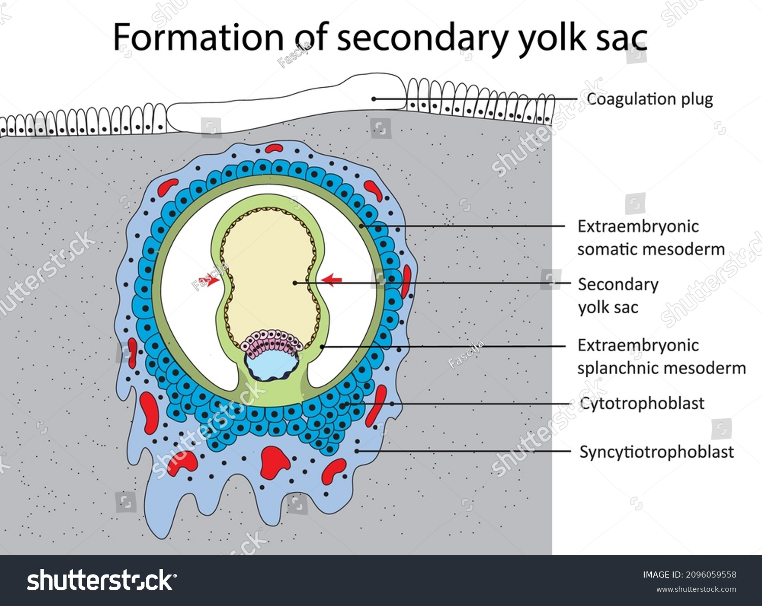
  • Secondary yolk sac also forms, when a large part of primary yolk sac separates and disappears leaving the smaller part. It is completely lined with endoderm.
Secondary Yolk Sac
  • Primary chorionic villi forms – cytotrophoblast projects into syncytiotrophoblast (more in the next chapter)

Syncytiotrophoblast – Placental barrier between maternal and fetal blood

Menstruation cycle and 1st week

menstruation cycle

Ovarian Cycle: Follicular phase – Ovulation – Luteal phase

Uterine Cycle: Menstruation – Proliferation – Secretory

Image result for menstruation cycle hormones

1. Follicle Stimulation Hormone (FSH)

  • Produced by anterior pituitary gland
  • Stimulates primary follicles to develop

2. Estrogen

  • Produced by primary follicles
  • Endometrium to proliferate
  • Stop production of FSH so that other primary follicle don’t develop
  • Stimulate production of LH (Luteinizing Hormone)

3. Luteinizing Hormone (LH)

  • Produced by anterior pituitary gland
  • Stimulates ovulation
  • Rest of Graffian follicle forms corpus luteum – produces progesterone

4. Progesterone

  • Maintain thick endometrium (secretory phase of endometrium)

NB:

  • If fertilized, corpus luteum will form corpus luteum of pregnancy – produces progesterone and is maintained by gonadotropic hormones produced by the embryo until placenta takes over to produce progesterone
  • If not fertilized, corpus luteum will form corpus albicans which will then degenerate
  • Reduction in production of progesterone leads to menstruation
  • Reduction in production of estrogen leads to FSH production
  • Proteolytic enzymes prevent coagulation of blood during menstruation
  1. Germinal period – 2 weeks; formation of 3 germ layers
  2. Embryonic period – 3rd week to 3rd month; differentiation of the 3 germ layers into organs and systems
  3. Fetal period – growth of various organs and systems

1st week after fertilization

Zygote →Cleavage division → Morula → Blastocyst → Hatching (degeneration of zona pellucida) → Late blastocyst

Image result for 1st week after fertilization

Inner cell mass forms embryoblast

Outer cell mass forms trophoblast; which has 2 poles:

  • Embryonic pole – faces site of implantation
  • Abembryonic pole – opposite pole

Image result for embryonic pole and abembryonic pole
blastocyst

Process of Implantation:

  1. Trophoblast erodes endometrium
  2. Penetration defect in endometrium
  3. Blastocyst embedded in endometrium of uterus
  4. Penetration defect closed by fibrin clot

Clinicals:

  1. Ectopic Pregnancy – implantation can occur in the abdomen, ovarian and fallopian tube
  2. Blighted ovum – dead embryoblast
  3. Downs syndrome – Trisomy of chromosome 21
  4. Turners syndrome – monosomy of x chromosome
  5. Triple x syndrome

Fertilization in mammals

  • Fusion of sperm nucleus with egg nucleus to form a diploid zygote cell
  • Occurs in upper parts of fallopian tube ie. Ampulla

Image result for ampulla of fallopian tube
parts of fallopian tube

Process of Fertilization

1. Ejaculation in vagina

2. Capacitation – removal of glycoproteins to make sperm active and identify ovum

3. 1st contact – Acrosome reaction

  • Contact of head of sperm with zona granulosa cells
  • Acrosome reaction – Acrosome burst by exocytosis
  • Releases hydrolytic enzymes

4. Break down of first barrier – Corona radiata

  • Enzymes digest the sticky substances that holds the follicle cells together
  • Cause cells to break away from zona pellucida

5. Break down of 2nd barrier – Zona pellucida

  • Another acrosomal enzyme then digests the zona pellucida to create path for entry of the sperm cell

6. Contact with third barrier – Oocyte membrane

Formation of fertilization membrane:

  • Head of 1st sperm makes contact with oocyte membrane
  • Oocyte membrane become permeable to calcium ions
  • Calcium ions rush into oocyte by diffusion
  • Calcium ions stimulate rapid exocytosis of cortical granules, which release chemical substances
  • Cortical reaction – release of cortical granules
  • A new membrane forms, called fertilization membrane
  • Fertilization membrane prevents polyspermy (entry of many sperm cells)

Q) How does fertilization membrane prevent polyspermy?

Acrosomal enzymes released by other sperms cannot digest the changed zona pellucida/fertilization membrane

7. Release of sperm nucleus into the secondary oocyte

8.  This stimulates 2ry oocyte to complete 2nd meiotic division and form a true ovum

9. Fusion of male and female pronucleus (enlarged nucleus) to form a zygote

Image result for corona radiata oocyte
Process of fertilization

Definition of terms

Embryology: study of developmental process from a single cell to a baby in months

Embryogenesis: formation and development of an embryo

Ontogeny: Process of development of an organism

Conceptus: Product of conception

Cleavage: series of mitotic cell divisions of the zygote resulting in blastomeres

Morula: a mass of 12 to 32 blastomeres

After a while, the morula develops a cavity and we call it a;

Blastocyst: structure formed after a cavity forms within the morula.

Blastocele: fluid that enters the cavity of blastocyst

Blastocele

Embryoblast: inner cell mass of blastocyst

Trophoblast: outer cell mass of blastocyst

Endometrium: inner epithelial lining of the uterus

Implantation: attachment and embedding of the blastocyst to the endometrium

Decidua: endometrium during pregnancy

Bilaminar disc: 2 layered embryo, occurs during second the 2nd week

Gastrula: trilaminar disc embryo (3 layered embryo)

ie. ectoderm, mesoderm, endoderm

Gastrulation: process of formation of the trilaminar disc, begins on 15th day

Neurulation: process of formation of neural tube, the primordium of the central nervous system

Primordium: beginning or first discernible indication of an organ or structure

Organogenesis: formation of body organs

Morphogenesis: process of shape development

Fetus: unborn offspring

Abortion: expulsion of an embryo or fetus before its viability (capable of living outside the uterus)

Teratology: study of birth defects

Teratogen: an agent that causes congenital (born with) defects

Anomaly: structural abnormality

Malformation: abnormal developmental process