Definitions
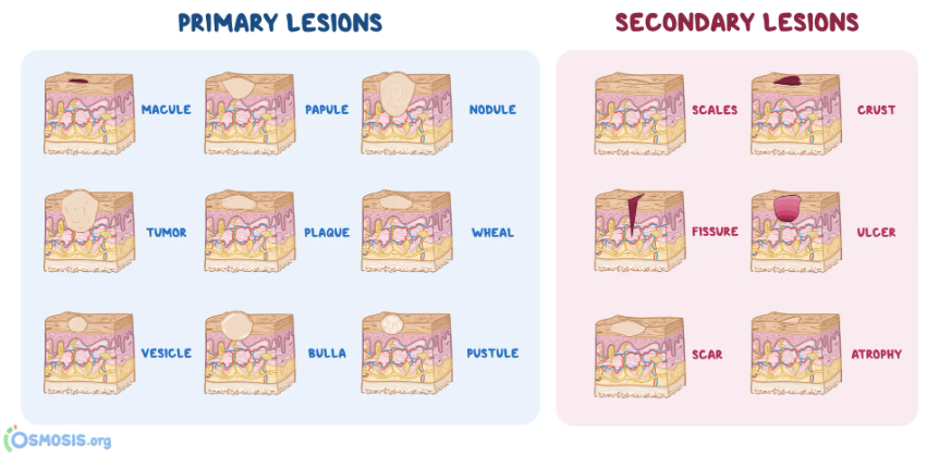
1. Macule
- Focal area of colour change
- No elevation or depression in relation to surroundings
2. Papule
- Solid raised lesion
- Less than 5mm in diameter
3. Nodule
- Solid raised lesion
- More than 5mm in diameter
4. Sessile
- Tumor/growth
- Base is widest part of lesion
5. Pedunculated
- Tumor/growth
- Base is narrower than widest part of lesion
6. Papillary
- Tumor/growth
- Numerous warty projections
7. Verrucous
- Tumor/growth
- Rough warty surface
8. Vesicle
- Superficial blister filled with clear fluid
- Less than 5mm in diameter
9. Bulla – Blister > 5mm in diameter
10. Pustule – Blister filled with purulent exudate
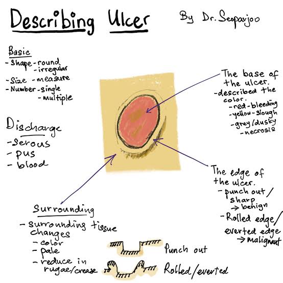
11. Ulcer
- Loss of surface epithelium ± underlying CT
- Depressed/ excavated
12. Erosion
- Superficial lesion
- Partial/ total loss of surface epithelium
- After rupture of vesicle or bulla
13. Fissure – Narrow slit like ulceration/ groove
14. Plaque – Elevated and flat on surface
15. Petechia – Round pinpoint area of hemorrhage
16. Ecchymosis – Non elevated hemorrhage, larger than petechia
17. Telangiectasia
- Vascular lesion
- Dilation of small superficial blood vessel
18. Cysts
- Pathological epithelium lined cavity
- Filled with liquid or semisolid content
19. Unilocular – Radiolucent lesion – single compartment
20. Multilocular – Radiolucent lesion – several compartments
Traumatic lesions
Clinical manifestations of trauma
- Acute/chronic ulcers
- Red/white lesions
- Mucositis
- Reactive hyperplasia
- Bone exposure and sequestration
Etiology
Physical trauma
- Factitial injury – Self induced/psychological
- Eg. Cheek biting – subconscious reaction to stress, emotions, boredom
- Riga-Fede disease – Traumatic ulcer on anterior tongue with natal teeth in infants, or repetitive tongue thrusting habit after eruption of 1ry lower incisors
- Frictional hyperkeratosis – white lesion, ill fitting dentures
- Iatrogenic
Therapeutic radiation
Classification
1. Acute traumatic ulcer
- Pain
- Yellow base, red halo
- Heals in 7 to 10 days
2. Chronic traumatic lesions
- Little or no pain
- Scar formation
- Mimic carcinoma/infective ulcer
Clinical
- Single lesion
- Erythematous
- Non inverted margins
- Clean base covered in pseudomembrane
- Painful
- Disappear in 7-10 days after eliminating cause
Histology
- Loss of epithelium – replaced by fibrin network
- Granulation tissue base
- Scar formation deep in tissues
- Dense inflammatory infiltrate – macrophages and eosinophils
Management
- Observe for 2 weeks to rule out infection
- Topical corticosteroids
Pathological lesions
Etiology
1. Neoplasm
2. Immunological disease
3. Aphthous ulcer/ Canker sore – Multifactorial
4. Infections
- Bacteria:
- Fungi
Nb: Chronic infectious ulcers (TB, syphilis, fungal)
- Mimic carcinoma/traumatic ulcer
- Non healing and persistent
- Multiple
- Diagnosis – Biopsy and culture
- Management – Antimicrobial agent
